 Explore
 Validate
 Learn
Learn Western blot
 ELISA
ELISAAntibody data
- Antibody Data
- Antigen structure
- References [2]
- Comments [0]
- Validations
- Western blot [2]
- Other assay [1]
Submit
Validation data
Reference
Comment
Report error
- Product number
- 37-0800 - Provider product page

- Provider
- Invitrogen Antibodies
- Product name
- KPNA1 Monoclonal Antibody (114-E12)
- Antibody type
- Monoclonal
- Antigen
- Other
- Reactivity
- Human, Mouse
- Host
- Mouse
- Isotype
- IgG
- Antibody clone number
- 114-E12
- Vial size
- 100 µg
- Concentration
- 0.5 mg/mL
- Storage
- -20°C
Submitted references Cell-to-cell transmission of C9orf72 poly-(Gly-Ala) triggers key features of ALS/FTD.
Assembly of the human origin recognition complex occurs through independent nuclear localization of its components.
Khosravi B, LaClair KD, Riemenschneider H, Zhou Q, Frottin F, Mareljic N, Czuppa M, Farny D, Hartmann H, Michaelsen M, Arzberger T, Hartl FU, Hipp MS, Edbauer D
The EMBO journal 2020 Apr 15;39(8):e102811
The EMBO journal 2020 Apr 15;39(8):e102811
Assembly of the human origin recognition complex occurs through independent nuclear localization of its components.
Ghosh S, Vassilev AP, Zhang J, Zhao Y, DePamphilis ML
The Journal of biological chemistry 2011 Jul 8;286(27):23831-41
The Journal of biological chemistry 2011 Jul 8;286(27):23831-41
No comments: Submit comment
Supportive validation
- Submitted by
- Invitrogen Antibodies (provider)
- Main image

- Experimental details
- Western blot analysis was performed on whole cell extracts (30 µg lysate) of HEL 92.1.7 (Lane 1), K-562 (Lane 2), U-2 OS (Lane 3), Caco-2 (Lane 4) and PC-3 (Lane 5). The blot was probed with Anti-NPI 1 Mouse Monoclonal Antibody (Product # 37-0800, 2 µg/mL) and detected by chemiluminescence using Goat anti-Mouse IgG (H+L) Superclonal™ Secondary Antibody, HRP conjµgate (Product # A28177, 0.4 µg/mL, 1:2500 dilution). A ~60 kDa band corresponding to NPI 1 was observed across the cell lines tested. Known quantity of protein samples were electrophoresed using Novex® NuPAGE® 4-12 % Bis-Tris gel (Product # NP0321BOX), XCell SureLock™ Electrophoresis System (Product # EI0002) and Novex® Sharp Pre-Stained Protein Standard (Product # LC5800). Resolved proteins were then transferred onto a nitrocellulose membrane with iBlot® 2 Dry Blotting System (Product # IB21001). The membrane was probed with the relevant primary and secondary Antibody using iBind™ Flex Western Starter Kit (Product # SLF2000S). Chemiluminescent detection was performed using Pierce™ ECL Western Blotting Substrate (Product # 32106).

- Submitted by
- Invitrogen Antibodies (provider)
- Main image

- Experimental details
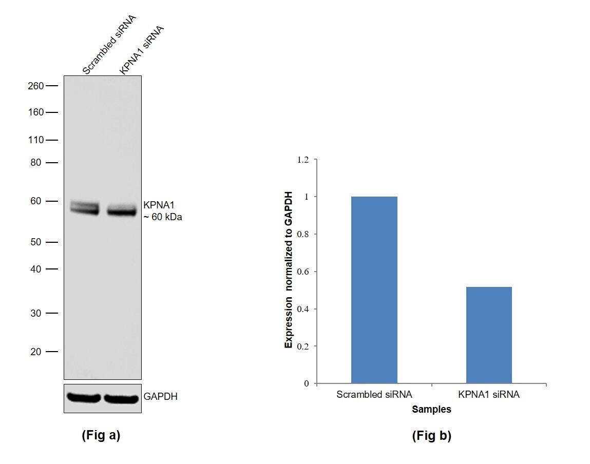
- Knockdown of KPNA1 was achieved by transfecting PC-3 with KPNA1 specific siRNAs (Silencer® select Product # S7915, S223979). Western blot analysis (Fig. a) was performed using Whole cell extracts from the KPNA1 knockdown cells (lane 2) and non-targeting scrambled siRNA transfected cells (lane 1). The blot was probed with KPNA1 Monoclonal Antibody (114-E12) (Product # 37-0800, 2 µg/mL ) and Goat anti-Mouse IgG (H+L) Superclonal™ Recombinant Secondary Antibody, HRP (Product # A28177, 1:4000). Densitometric analysis of this western blot for the upper band is shown in histogram (Fig. b). Although the lower band shows no reduction in signal, the decrease in signal for the upper band upon siRNA mediated knock down confirms that antibody is specific to KPNA1.
Supportive validation
- Submitted by
- Invitrogen Antibodies (provider)
- Main image
- Experimental details
- NULL
