 Explore
 Validate
 Learn
Learn Western blot
Antibody data
- Antibody Data
- Antigen structure
- References [0]
- Comments [0]
- Validations
- Western blot [2]
- Immunocytochemistry [1]
- Immunohistochemistry [6]
Submit
Validation data
Reference
Comment
Report error
- Product number
- AMAb90786 - Provider product page

- Provider
- Atlas Antibodies
- Proper citation
- Atlas Antibodies Cat#AMAb90786, RRID:AB_2665666
- Product name
- Anti-PHGDH
- Antibody type
- Monoclonal
- Reactivity
- Human
- Host
- Mouse
- Conjugate
- Unconjugated
- Antigen sequence
LEEIWPLCDFITVHTPLLPSTTGLLNDNTFAQCKK
GVRVVNCARGGIVDEGALLRALQSGQCAGAALDVF
TEEPPRDRALVDHENVISCPHLGASTKEAQSRCGE
EIAVQFVDM- Isotype
- IgG
- Antibody clone number
- CL0555
- Vial size
- 100 µl
- Storage
- Store at +4°C for short term storage. Long time storage is recommended at -20°C.
No comments: Submit comment
Supportive validation
- Submitted by
- Atlas Antibodies (provider)
- Main image

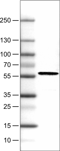
- Experimental details
- Lane 1: Marker [kDa]Lane 2: Human cell line RT-4
- Submitted by
- Atlas Antibodies (provider)
- Main image

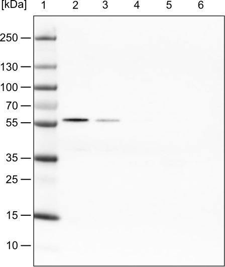
- Experimental details
- Lane 1: Marker [kDa]Lane 2: Human cell line HeLa cytoplasmic fractionLane 3: Human cell line HeLa membrane fractionLane 4: Human cell line HeLa nuclear fractionLane 5: Human cell line HeLa chromatin fractionLane 6: Human cell line HeLa cytoskeletal fraction
Supportive validation
- Submitted by
- Atlas Antibodies (provider)
- Main image

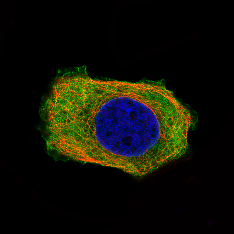
- Experimental details
- Immunofluorescence staining of MCF7 cells using the Anti-PHGDH monoclonal antibody, showing specific staining in the cytosol and the plasma membrane in green. Microtubule- and nuclear probes are visualized in red and blue, respectively (where available).
- Sample type
- HUMAN
Supportive validation
- Submitted by
- Atlas Antibodies (provider)
- Main image

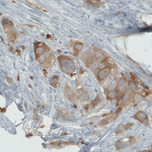
- Experimental details
- Immunohistochemical staining of human breast cancer shows moderate cytoplasmic immunoreactivity in tumour cells.
- Submitted by
- Atlas Antibodies (provider)
- Main image

- Experimental details
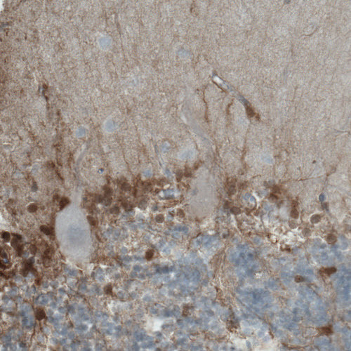
- Immunohistochemical staining of human cerebellum shows moderate positivity in the molecular cell layer (basket cells).
- Submitted by
- Atlas Antibodies (provider)
- Main image

- Experimental details
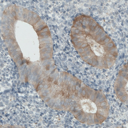
- Immunohistochemical staining of human uterus shows moderate cytoplasmic immunoreactivity in the glandular epithelium cells.
- Submitted by
- Atlas Antibodies (provider)
- Main image

- Experimental details
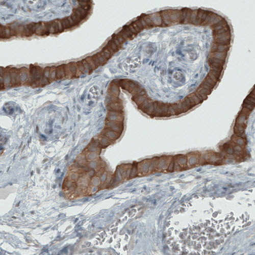
- Immunohistochemical staining of human fallopian tube shows strong immunoreactivity is the epithelial cells.
- Submitted by
- Atlas Antibodies (provider)
- Main image

- Experimental details
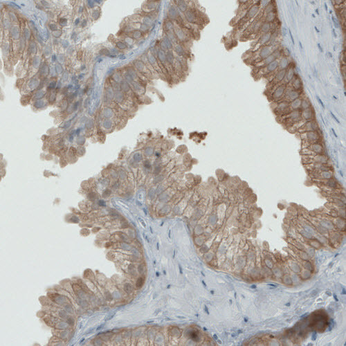
- Immunohistochemical staining of human prostate shows moderate positivity in the glandular cells.
- Submitted by
- Atlas Antibodies (provider)
- Main image
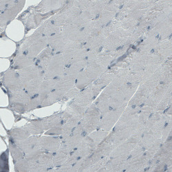
- Experimental details
- Immunohistochemical staining of human striated muscle shows absence of immunoreactivity (negative control).
