Explore
 Validate
 Learn
Learn Western blot
 ELISA
ELISAAntibody data
- Antibody Data
- Antigen structure
- References [4]
- Comments [0]
- Validations
- Western blot [1]
- Immunocytochemistry [1]
Submit
Validation data
Reference
Comment
Report error
- Product number
- 14821-1-AP - Provider product page

- Provider
- Proteintech Group
- Proper citation
- Proteintech Cat#14821-1-AP, RRID:AB_2219204
- Product name
- SARA antibody
- Antibody type
- Polyclonal
- Description
- KD/KO validated SARA antibody (Cat. #14821-1-AP) is a rabbit polyclonal antibody that shows reactivity with human, mouse and has been validated for the following applications: ELISA.
- Reactivity
- Human, Mouse
- Host
- Rabbit
- Conjugate
- Unconjugated
- Isotype
- IgG
- Vial size
- 20ul, 150ul
Submitted references WDR81 regulates adult hippocampal neurogenesis through endosomal SARA-TGFβ signaling.
Dachshund Depletion Disrupts Mammary Gland Development and Diverts the Composition of the Mammary Gland Progenitor Pool.
TGFBR-IDH1-Cav1 axis promotes TGF-β signalling in cancer-associated fibroblast.
Phosphatidylinositol 3-kinase and Rab5 GTPase inversely regulate the Smad anchor for receptor activation (SARA) protein independently of transforming growth factor-β1.
Wang M, Tang C, Xing R, Liu X, Han X, Liu Y, Wang L, Yang C, Guo W
Molecular psychiatry 2021 Feb;26(2):694-709
Molecular psychiatry 2021 Feb;26(2):694-709
Dachshund Depletion Disrupts Mammary Gland Development and Diverts the Composition of the Mammary Gland Progenitor Pool.
Jiao X, Li Z, Wang M, Katiyar S, Di Sante G, Farshchian M, South AP, Cocola C, Colombo D, Reinbold R, Zucchi I, Wu K, Tabas I, Spike BT, Pestell RG
Stem cell reports 2019 Jan 8;12(1):135-151
Stem cell reports 2019 Jan 8;12(1):135-151
TGFBR-IDH1-Cav1 axis promotes TGF-β signalling in cancer-associated fibroblast.
Hou X, Zhang J, Wang Y, Xiong W, Mi J
Oncotarget 2017 Oct 13;8(48):83962-83974
Oncotarget 2017 Oct 13;8(48):83962-83974
Phosphatidylinositol 3-kinase and Rab5 GTPase inversely regulate the Smad anchor for receptor activation (SARA) protein independently of transforming growth factor-β1.
Runyan CE, Liu Z, Schnaper HW
The Journal of biological chemistry 2012 Oct 19;287(43):35815-24
The Journal of biological chemistry 2012 Oct 19;287(43):35815-24
No comments: Submit comment
Supportive validation
- Submitted by
- Proteintech Group (provider)
- Main image
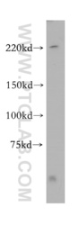
- Experimental details
- mouse brain tissue were subjected to SDS PAGE followed by western blot with 14821-1-AP(ZFYVE9 antibody) at dilution of 1:1000
- Sample type
- tissue

Supportive validation
- Submitted by
- Proteintech Group (provider)
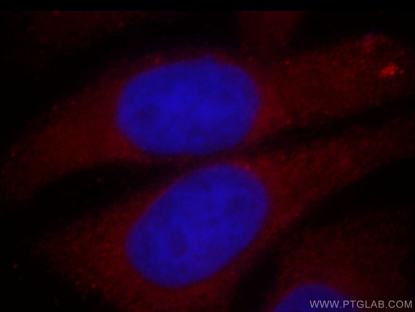
- Main image
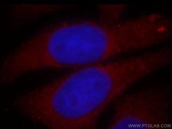
- Experimental details
- Immunofluorescent analysis of HepG2 cells, using ZFYVE9 antibody 14821-1-AP at 1:25 dilution and Rhodamine-labeled goat anti-rabbit IgG (red). Blue pseudocolor = DAPI (fluorescent DNA dye).
- Sample type
- cell line