Explore
 Validate
 Learn
Learn Western blot
Antibody data
- Antibody Data
- Antigen structure
- References [1]
- Comments [0]
- Validations
- Western blot [1]
- Immunocytochemistry [1]
Submit
Validation data
Reference
Comment
Report error
- Product number
- AF4939 - Provider product page

- Provider
- R&D Systems
- Product name
- Anti-Human MST1/STK4 Antigen Affinity-purified Polyclonal Antibody
- Antibody type
- Polyclonal
- Antigen
- E. coli-derived recombinant human MST1/STK4, Lys285-Asp443
- Description
- Antigen Affinity-purified
- Reactivity
- Human
- Host
- Goat
- Antigen sequence
Q13043- Isotype
- IgG
- Vial size
- 100 µg
Submitted references Identification of microRNA signature in the progression of gestational trophoblastic disease.
Zhao JR, Cheng WW, Wang YX, Cai M, Wu WB, Zhang HJ
Cell death & disease 2018 Jan 24;9(2):94
Cell death & disease 2018 Jan 24;9(2):94
No comments: Submit comment
Supportive validation
- Submitted by
- R&D Systems (provider)
- Main image

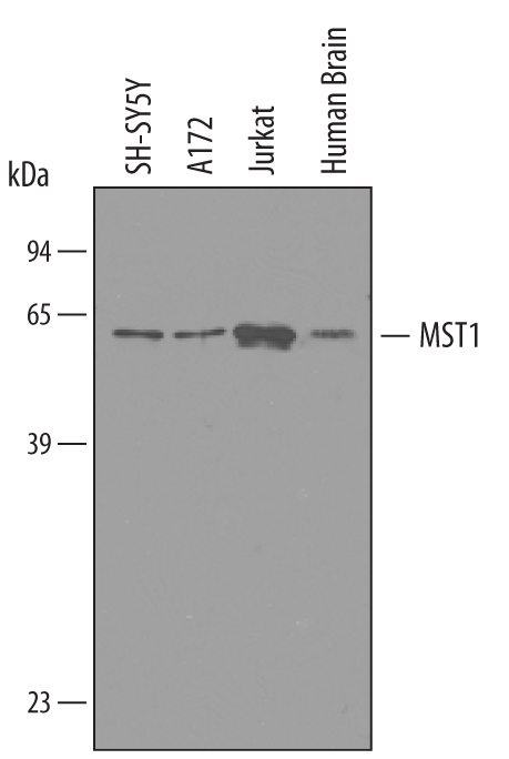
- Experimental details
- Detection of Human MST1/STK4 by Western Blot. Western blot shows lysates of human brain tissue, A172 human glioblastoma cell line, Jurkat human acute T cell leukemia cell line, and SH-SY5Y human neuroblastoma cell line. PVDF membrane was probed with 1 µg/mL of Goat Anti-Human MST1/STK4 Antigen Affinity-purified Polyclonal Antibody (Catalog # AF4939) followed by HRP-conjugated Anti-Goat IgG Secondary Antibody (Catalog # HAF019). A specific band was detected for MST1/STK4 at approximately 55-60 kDa (as indicated). This experiment was conducted under reducing conditions and using Immunoblot Buffer Group 8.
Supportive validation
- Submitted by
- R&D Systems (provider)
- Main image

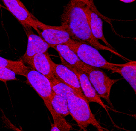
- Experimental details
- MST1/STK4 in SH-SY5Y Human Cell Line. MST1/STK4 was detected in immersion fixed SH-SY5Y human neuroblastoma cell line using Goat Anti-Human MST1/STK4 Antigen Affinity-purified Polyclonal Antibody (Catalog # AF4939) at 10 µg/mL for 3 hours at room temperature. Cells were stained using the NorthernLights™ 557-conjugated Anti-Goat IgG Secondary Antibody (red; Catalog # NL001) and counterstained with DAPI (blue). Specific staining was localized to cytoplasm. View our protocol for Fluorescent ICC Staining of Cells on Coverslips.