Explore
 Validate
 Learn
Learn Western blot
Antibody data
- Antibody Data
- Antigen structure
- References [6]
- Comments [0]
- Validations
- Western blot [1]
- Immunohistochemistry [1]
- Flow cytometry [1]
- Blocking/Neutralizing [1]
Submit
Validation data
Reference
Comment
Report error
- Product number
- AF4160 - Provider product page

- Provider
- R&D Systems
- Product name
- Human Plexin D1 Antibody
- Antibody type
- Polyclonal
- Description
- Antigen Affinity-purified. Detects human Plexin D1 in direct ELISAs and Western blots.
- Reactivity
- Human
- Host
- Goat
- Conjugate
- Unconjugated
- Antigen sequence
Q9Y4D7- Isotype
- IgG
- Vial size
- 100 ug
- Concentration
- LYOPH
- Storage
- Use a manual defrost freezer and avoid repeated freeze-thaw cycles. 12 months from date of receipt, -20 to -70 °C as supplied. 1 month, 2 to 8 °C under sterile conditions after reconstitution. 6 months, -20 to -70 °C under sterile conditions after reconstitution.
Submitted references Post-endocytic sorting of Plexin-D1 controls signal transduction and development of axonal and vascular circuits.
Chemorepellent Semaphorin 3E Negatively Regulates Neutrophil Migration In Vitro and In Vivo.
Semaphorin 3G Provides a Repulsive Guidance Cue to Lymphatic Endothelial Cells via Neuropilin-2/PlexinD1.
An image-based RNAi screen identifies SH3BP1 as a key effector of Semaphorin 3E-PlexinD1 signaling.
Dual role for Islet-1 in promoting striatonigral and repressing striatopallidal genetic programs to specify striatonigral cell identity.
Sema3E-Plexin D1 signaling drives human cancer cell invasiveness and metastatic spreading in mice.
Burk K, Mire E, Bellon A, Hocine M, Guillot J, Moraes F, Yoshida Y, Simons M, Chauvet S, Mann F
Nature communications 2017 Feb 22;8:14508
Nature communications 2017 Feb 22;8:14508
Chemorepellent Semaphorin 3E Negatively Regulates Neutrophil Migration In Vitro and In Vivo.
Movassagh H, Saati A, Nandagopal S, Mohammed A, Tatari N, Shan L, Duke-Cohan JS, Fowke KR, Lin F, Gounni AS
Journal of immunology (Baltimore, Md. : 1950) 2017 Feb 1;198(3):1023-1033
Journal of immunology (Baltimore, Md. : 1950) 2017 Feb 1;198(3):1023-1033
Semaphorin 3G Provides a Repulsive Guidance Cue to Lymphatic Endothelial Cells via Neuropilin-2/PlexinD1.
Liu X, Uemura A, Fukushima Y, Yoshida Y, Hirashima M
Cell reports 2016 Nov 22;17(9):2299-2311
Cell reports 2016 Nov 22;17(9):2299-2311
An image-based RNAi screen identifies SH3BP1 as a key effector of Semaphorin 3E-PlexinD1 signaling.
Tata A, Stoppel DC, Hong S, Ben-Zvi A, Xie T, Gu C
The Journal of cell biology 2014 May 26;205(4):573-90
The Journal of cell biology 2014 May 26;205(4):573-90
Dual role for Islet-1 in promoting striatonigral and repressing striatopallidal genetic programs to specify striatonigral cell identity.
Lu KM, Evans SM, Hirano S, Liu FC
Proceedings of the National Academy of Sciences of the United States of America 2014 Jan 7;111(1):E168-77
Proceedings of the National Academy of Sciences of the United States of America 2014 Jan 7;111(1):E168-77
Sema3E-Plexin D1 signaling drives human cancer cell invasiveness and metastatic spreading in mice.
Casazza A, Finisguerra V, Capparuccia L, Camperi A, Swiercz JM, Rizzolio S, Rolny C, Christensen C, Bertotti A, Sarotto I, Risio M, Trusolino L, Weitz J, Schneider M, Mazzone M, Comoglio PM, Tamagnone L
The Journal of clinical investigation 2010 Aug;120(8):2684-98
The Journal of clinical investigation 2010 Aug;120(8):2684-98
No comments: Submit comment
Supportive validation
- Submitted by
- R&D Systems (provider)
- Main image

- Experimental details
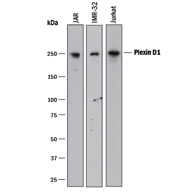
- Detection of Human Plexin D1 by Western Blot. Western blot shows lysates of JAR human choriocarcinoma cell line, IMR-32 human neuroblastoma cell line, and Jurkat human acute T cell leukemia cell line. PVDF membrane was probed with 0.5 µg/mL of Goat Anti-Human Plexin D1 Antigen Affinity-purified Polyclonal Antibody (Catalog # AF4160) followed by HRP-conjugated Anti-Goat IgG Secondary Antibody (Catalog # HAF017). A specific band was detected for Plexin D1 at approximately 250 kDa (as indicated). This experiment was conducted under reducing conditions and using Immunoblot Buffer Group 1.
Supportive validation
- Submitted by
- R&D Systems (provider)
- Main image

- Experimental details
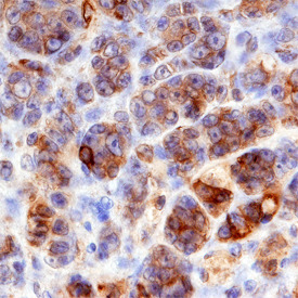
- Plexin D1 in Human Melanoma. Plexin D1 was detected in immersion fixed paraffin-embedded sections of human melanoma using Goat Anti-Human Plexin D1 Antigen Affinity-purified Polyclonal Antibody (Catalog # AF4160) at 3 µg/mL overnight at 4 °C. Before incubation with the primary antibody, tissue was subjected to heat-induced epitope retrieval using Antigen Retrieval Reagent-Basic (Catalog # CTS013). Tissue was stained using the Anti-Goat HRP-DAB Cell & Tissue Staining Kit (brown; Catalog # CTS008) and counterstained with hematoxylin (blue). Specific staining was localized to plasma membranes. View our protocol for Chromogenic IHC Staining of Paraffin-embedded Tissue Sections.
Supportive validation
- Submitted by
- R&D Systems (provider)
- Main image
- Experimental details
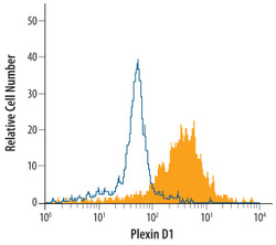
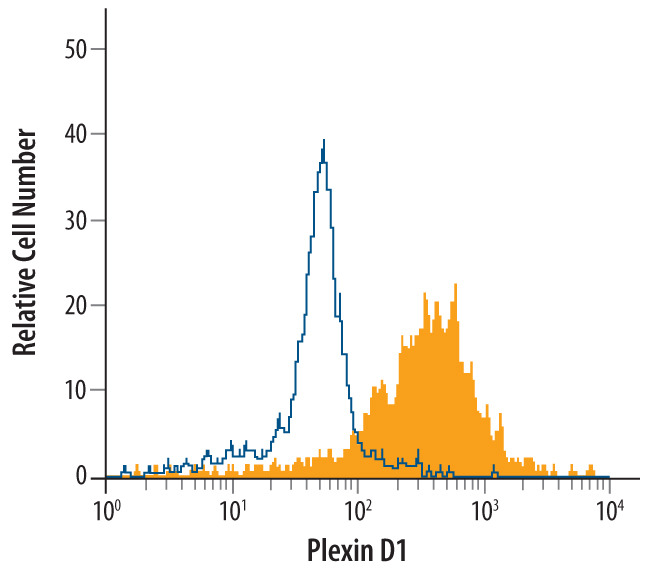
- Detection of Plexin D1 in Human peripheral blood monocytes by Flow Cytometry. Human peripheral blood monocytes were stained with Goat Anti-Human Plexin D1 Antigen Affinity-purified Polyclonal Antibody (Catalog # AF4160, filled histogram) or control antibody (Catalog # AB-108-C, open histogram), followed by Phycoerythrin-conjugated Anti-Goat IgG Secondary Antibody (Catalog # F0107).
Supportive validation
- Submitted by
- R&D Systems (provider)
- Main image

- Experimental details
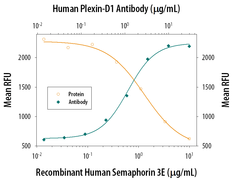
- Proliferation Inhibited by Semaphorin 3E and Neutralization by Human Plexin D1 Antibody. Recombinant Human Semaphorin 3E (Catalog # 3239-S3) inhibits proliferation in the HUVEC human umbilical vein endothelial cells in a dose-dependent manner (orange line). Plexin D1 mediated inhibition elicited by Recombinant Human Semaphorin 3E (5 ug/mL) is neutralized (green line) by increasing concentrations of Goat Anti-Human Plexin D1 Antigen Affinity-purified Polyclonal Antibody (Catalog # AF4160). The ND50 is typically 0.5-2 ug/mL.