 Explore
 Validate
 Learn
Learn Western blot
Antibody data
- Antibody Data
- Antigen structure
- References [4]
- Comments [0]
- Validations
- Western blot [1]
- Immunohistochemistry [1]
- Flow cytometry [1]
Submit
Validation data
Reference
Comment
Report error
- Product number
- MAB6285 - Provider product page

- Provider
- Novus Biologicals
- Product name
- Mouse Monoclonal ULBP-4/RAET1E Antibody
- Antibody type
- Monoclonal
- Description
- Protein A or G purified from hybridoma culture supernatant. Detects human ULBP-4/RAET1E in direct ELISAs and Western blots. In Western blots, approximately 20% cross-reactivity with recombinant human (rh) ULBP-2 is observed and no cross-reactivity with rhULBP-1 or rhULBP-3 is observed.
- Reactivity
- Human
- Host
- Mouse
- Conjugate
- Unconjugated
- Isotype
- IgG
- Vial size
- 100 ug
- Concentration
- LYOPH
- Storage
- Use a manual defrost freezer and avoid repeated freeze-thaw cycles. 12 months from date of receipt, -20 to -70 degreesC as supplied. 1 month, 2 to 8 degreesC under sterile conditions after reconstitution. 6 months, -20 to -70 degreesC under sterile conditions after reconstitution.
Submitted references Clinicopathological relevance of tumor expression of NK group 2 member D ligands in resected non-small cell lung cancer.
Targeting chemotherapy-resistant leukemia by combining DNT cellular therapy with conventional chemotherapy.
Adoptively transferred Vγ9Vδ2 T cells show potent antitumor effects in a preclinical B cell lymphomagenesis model.
MicroRNA-519a-3p mediates apoptosis resistance in breast cancer cells and their escape from recognition by natural killer cells.
Okita R, Maeda A, Shimizu K, Nojima Y, Saisho S, Nakata M
Oncotarget 2019 Nov 26;10(63):6805-6815
Oncotarget 2019 Nov 26;10(63):6805-6815
Targeting chemotherapy-resistant leukemia by combining DNT cellular therapy with conventional chemotherapy.
Chen B, Lee JB, Kang H, Minden MD, Zhang L
Journal of experimental & clinical cancer research : CR 2018 Apr 24;37(1):88
Journal of experimental & clinical cancer research : CR 2018 Apr 24;37(1):88
Adoptively transferred Vγ9Vδ2 T cells show potent antitumor effects in a preclinical B cell lymphomagenesis model.
Zumwalde NA, Sharma A, Xu X, Ma S, Schneider CL, Romero-Masters JC, Hudson AW, Gendron-Fitzpatrick A, Kenney SC, Gumperz JE
JCI insight 2017 Jul 6;2(13)
JCI insight 2017 Jul 6;2(13)
MicroRNA-519a-3p mediates apoptosis resistance in breast cancer cells and their escape from recognition by natural killer cells.
Breunig C, Pahl J, Küblbeck M, Miller M, Antonelli D, Erdem N, Wirth C, Will R, Bott A, Cerwenka A, Wiemann S
Cell death & disease 2017 Aug 3;8(8):e2973
Cell death & disease 2017 Aug 3;8(8):e2973
No comments: Submit comment
Supportive validation
- Submitted by
- Novus Biologicals (provider)
- Main image

- Experimental details
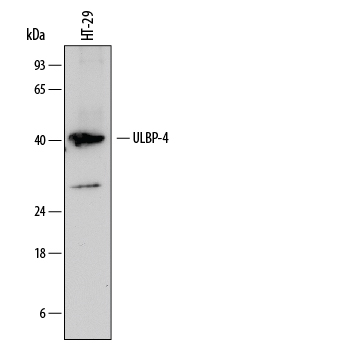
- Detection of Human ULBP-4/RAET1E by Western Blot. Western blot shows lysates of HT-29 human colon adenocarcinoma cell line. PVDF membrane was probed with 1 µg/mL of Mouse Anti-Human ULBP-4/ RAET1E Monoclonal Antibody (Catalog # MAB6285) followed by HRP-conjugated Anti-Mouse IgG Secondary Antibody (Catalog # HAF007). A specific band was detected for ULBP-4/RAET1E at approximately 40 kDa (as indicated). This experiment was conducted under reducing conditions and using Immunoblot Buffer Group 1.
Supportive validation
- Submitted by
- Novus Biologicals (provider)
- Main image

- Experimental details
- ULBP-4/RAET1E in Human Ovarian Cancer Tissue. ULBP-4/RAET1E was detected in immersion fixed paraffin-embedded sections of human ovarian cancer tissue using Mouse Anti-Human ULBP-4/ RAET1E Monoclonal Antibody (Catalog # MAB6285) at 15 µg/mL overnight at 4 °C. Tissue was stained using the Anti-Mouse HRP-DAB Cell & Tissue Staining Kit (brown; Catalog # CTS002) and counter-stained with hematoxylin (blue). Specific staining was localized to plasma membranes of epithelial cells. View our protocol for Chromogenic IHC Staining of Paraffin-embedded Tissue Sections.

Supportive validation
- Submitted by
- Novus Biologicals (provider)
- Main image

- Experimental details
- Detection of ULBP-4/RAET1E in HepG2 Human Cell Line by Flow Cytometry. HepG2 human hepatocellular carcinoma cell line was stained with Mouse Anti-Human ULBP-4/RAET1E Monoclonal Antibody (Catalog # MAB6285, filled histogram) or isotype control antibody (Catalog # MAB0041, open histogram), followed by Allophycocyanin-conjugated Anti-Mouse IgG Secondary Antibody (Catalog # F0101B).
