Explore
 Validate
 Learn
Learn Western blot
Antibody data
- Antibody Data
- Antigen structure
- References [1]
- Comments [0]
- Validations
- Western blot [3]
- Immunocytochemistry [1]
Submit
Validation data
Reference
Comment
Report error
- Product number
- AF3329 - Provider product page

- Provider
- R&D Systems
- Product name
- Human/Mouse Serum Albumin Antibody
- Antibody type
- Polyclonal
- Description
- Antigen Affinity-purified. Detects human and mouse Serum Albumin in direct ELISAs and Western blots.
- Reactivity
- Human, Mouse
- Host
- Goat
- Conjugate
- Unconjugated
- Isotype
- IgG
- Vial size
- 100 ug
- Storage
- Use a manual defrost freezer and avoid repeated freeze-thaw cycles. 12 months from date of receipt, -20 to -70 °C as supplied. 1 month, 2 to 8 °C under sterile conditions after reconstitution. 6 months, -20 to -70 °C under sterile conditions after reconstitution.
Submitted references Opposing impacts on healthspan and longevity by limiting dietary selenium in telomere dysfunctional mice.
Wu RT, Cao L, Mattson E, Witwer KW, Cao J, Zeng H, He X, Combs GF Jr, Cheng WH
Aging cell 2017 Feb;16(1):125-135
Aging cell 2017 Feb;16(1):125-135
No comments: Submit comment
Supportive validation
- Submitted by
- R&D Systems (provider)
- Main image

- Experimental details
- Detection of Human and Mouse Serum Albumin by Western Blot. Western blot shows lysates of human liver tissue and mouse liver tissue. PVDF membrane was probed with 0.1 µg/mL of Goat Anti-Human/Mouse Serum Albumin Antigen Affinity-purified Polyclonal Antibody (Catalog # AF3329) followed by HRP-conjugated Anti-Goat IgG Secondary Antibody (Catalog # HAF017). A specific band was detected for Serum Albumin at approximately 65-70 kDa (as indicated). This experiment was conducted under reducing conditions and using Immunoblot Buffer Group 1.

- Submitted by
- R&D Systems (provider)
- Main image

- Experimental details
- Detection of Human and Mouse Albumin by Simple WesternTM. Simple Western lane view shows lysates of human liver tissue and mouse liver tissue, loaded at 0.2 mg/mL. A specific band was detected for Albumin at approximately 64 kDa (as indicated) using 10 µg/mL of Goat Anti-Human/Mouse Serum Albumin Antigen Affinity-purified Polyclonal Antibody (Catalog # AF3329) followed by 1:50 dilution of HRP-conjugated Anti-Goat IgG Secondary Antibody (Catalog # HAF109). This experiment was conducted under reducing conditions and using the 12-230 kDa separation system.

- Submitted by
- R&D Systems (provider)
- Main image

- Experimental details
- Detection of Human Albumin by Simple WesternTM. Simple Western lane view shows lysates of human serum, loaded at 1:25000. A specific band was detected for Albumin at approximately 64 kDa (as indicated) using 5 µg/mL of Goat Anti-Human/Mouse Serum Albumin Antigen Affinity-purified Polyclonal Antibody (Catalog # AF3329) followed by 1:50 dilution of HRP-conjugated Anti-Goat IgG Secondary Antibody (Catalog # HAF109). This experiment was conducted under reducing conditions and using the 12-230 kDa separation system.

Supportive validation
- Submitted by
- R&D Systems (provider)
- Main image

- Experimental details
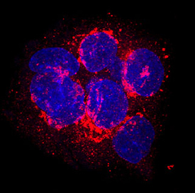
- Serum Albumin in HepG2 Human Cell Line. Serum Albumin was detected in immersion fixed HepG2 human hepatocellular carcinoma cell line using Goat Anti-Human/Mouse Serum Albumin Antigen Affinity-purified Polyclonal Antibody (Catalog # AF3329) at 10 µg/mL for 3 hours at room temperature. Cells were stained using the NorthernLights™ 557-conjugated Anti-Goat IgG Secondary Antibody (red; Catalog # NL001) and counterstained with DAPI (blue). Specific staining was localized to cytoplasm. View our protocol for Fluorescent ICC Staining of Cells on Coverslips.