Explore
 Validate
 Learn
Learn Western blot
 ELISA
ELISAAntibody data
- Antibody Data
- Antigen structure
- References [0]
- Comments [0]
- Validations
- Western blot [3]
- Immunocytochemistry [1]
- Immunohistochemistry [2]
Submit
Validation data
Reference
Comment
Report error
- Product number
- PA5-98458 - Provider product page

- Provider
- Invitrogen Antibodies
- Product name
- PLS1 Polyclonal Antibody
- Antibody type
- Polyclonal
- Antigen
- Recombinant full-length protein
- Reactivity
- Human
- Host
- Rabbit
- Isotype
- IgG
- Vial size
- 100 µg
- Concentration
- 1 mg/mL
- Storage
- -20°C or -80°C if preferred
No comments: Submit comment
Supportive validation
- Submitted by
- Invitrogen Antibodies (provider)
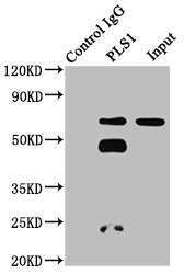
- Main image

- Experimental details
- Western Blot analysis of precipitated PLS1 from 293 whole cell lysate using a PLS1 polyclonal antibody (Product # PA5-98458). An HRP-conjugated Protein G antibody was used as the secondary antibody (1:2000). Lane 1: Rabbit control IgG. Lane 2: 293 whole cell lysate (500µg). Lane 3: 293 whole cell lysate (20µg).
- Submitted by
- Invitrogen Antibodies (provider)
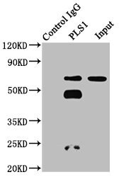
- Main image

- Experimental details
- Western Blot analysis of precipitated PLS1 from 293 whole cell lysate using a PLS1 polyclonal antibody (Product # PA5-98458). An HRP-conjugated Protein G antibody was used as the secondary antibody (1:2000). Lane 1: Rabbit control IgG. Lane 2: 293 whole cell lysate (500µg). Lane 3: 293 whole cell lysate (20µg).
- Submitted by
- Invitrogen Antibodies (provider)
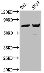
- Main image

- Experimental details
- Western Blot analysis of PLS1 using a PLS1 Polyclonal antibody (Product # PA5-98458) at a concentration of 5.2 µg/mL. Positive WB detected in: 293 whole cell lysate, A549 whole cell lysate. A secondary Goat polyclonal antibody to rabbit IgG was applied at a 1:50,000 dilution. Observed band size: 71 kDa.
Supportive validation
- Submitted by
- Invitrogen Antibodies (provider)
- Main image
- Experimental details
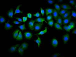
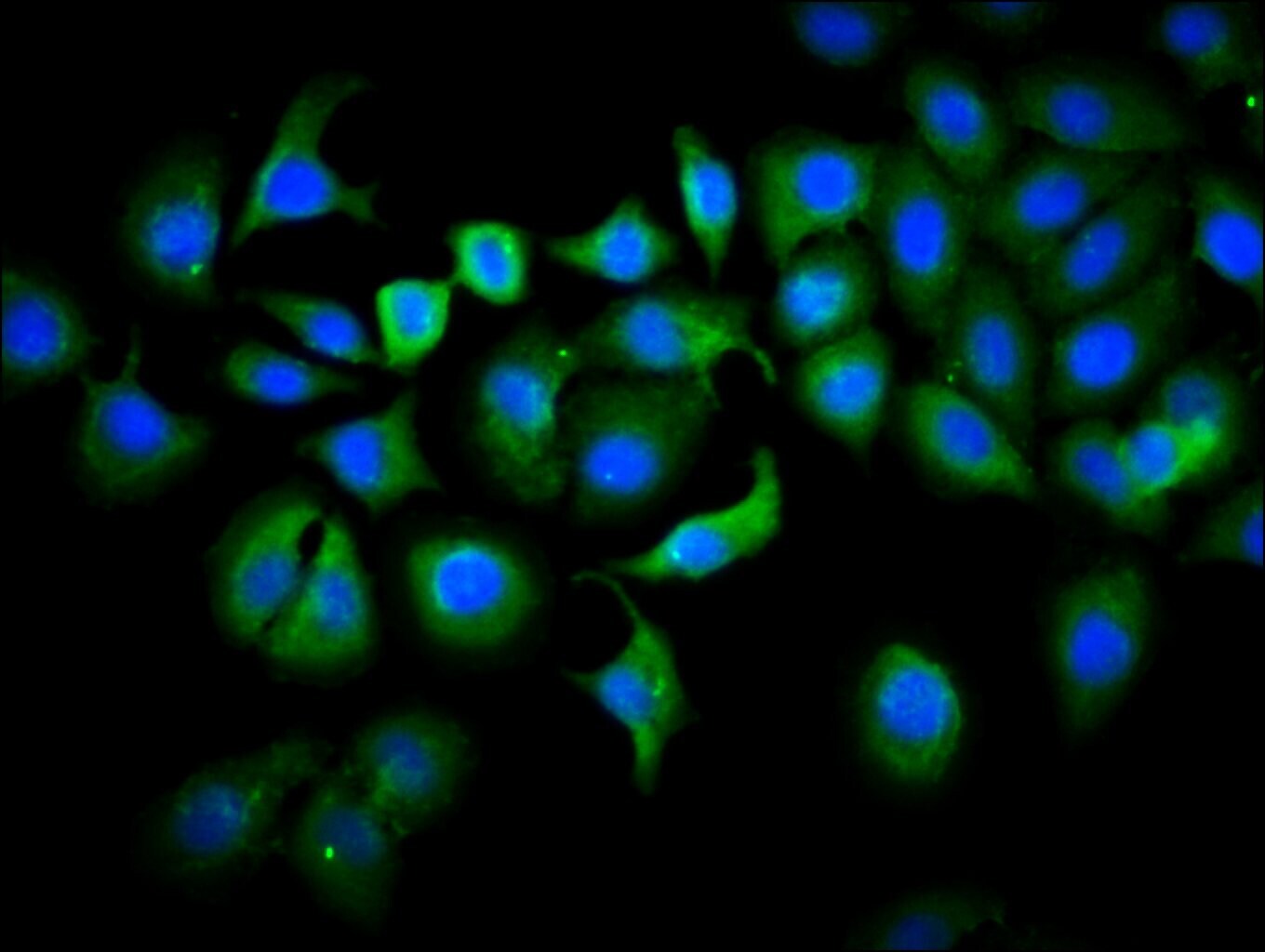
- Immunofluorescent analysis of PLS1 in A549 cells using a PLS1 polyclonal antibody (Product # PA5-98458) at a dilution of 1:133. The cells were fixed in 4% formaldehyde, permeabilized using 0.2% Triton X-100 and blocked in 10% normal Goat Serum. The cells were then incubated with the antibody overnight at 4°C. The secondary antibody was Alexa Fluor 488-congugated Goat Anti-Rabbit IgG(H+L). Cells were counter-stained with DAPI.
Supportive validation
- Submitted by
- Invitrogen Antibodies (provider)
- Main image
- Experimental details
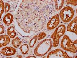
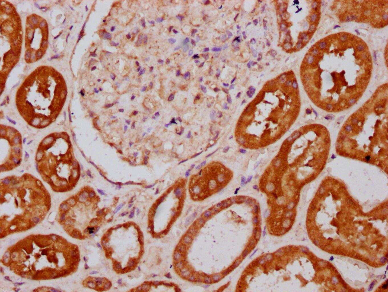
- Immunohistochemical analysis of PLS1 in paraffin embedded human kidney tissue using a PLS1 polyclonal antibody (Product # PA5-98458) at a dilution of 1:400. After dewaxing and hydration, antigen retrieval was mediated by high pressure in a citrate buffer (pH 6.0). Section was blocked with 10% normal goat serum 30min at RT. Then primary antibody (1% BSA) was incubated at 4°C overnight. The primary is detected by a biotinylated secondary antibody and visualized using an HRP conjugated SP system.
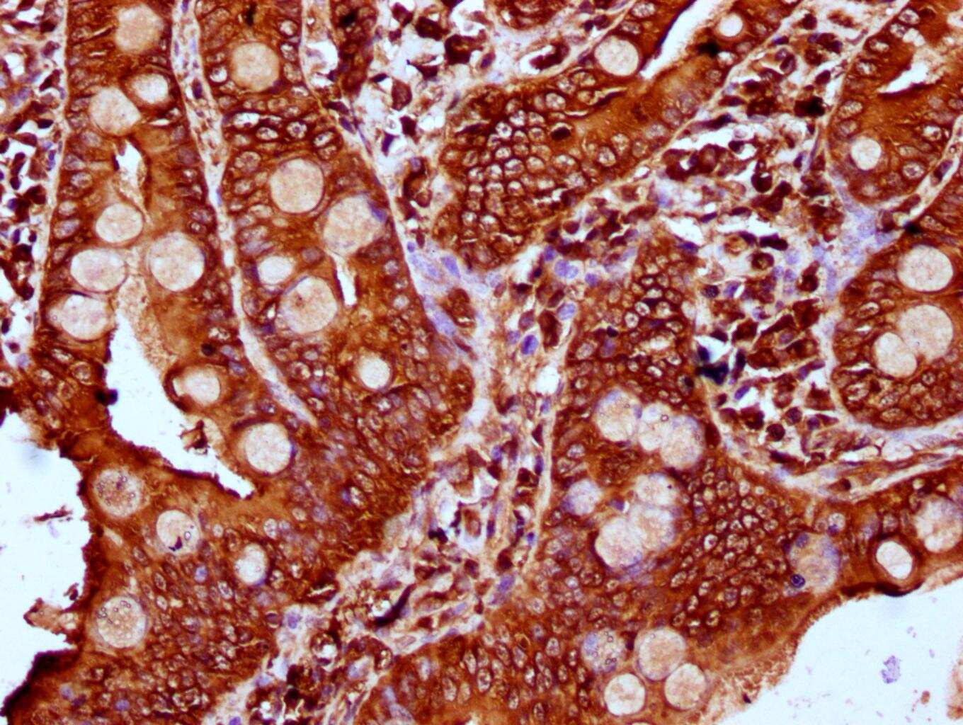
- Submitted by
- Invitrogen Antibodies (provider)
- Main image
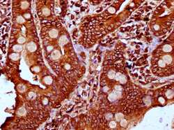
- Experimental details
- Immunohistochemical analysis of PLS1 in paraffin embedded human small intestine tissue using a PLS1 polyclonal antibody (Product # PA5-98458) at a dilution of 1:400. After dewaxing and hydration, antigen retrieval was mediated by high pressure in a citrate buffer (pH 6.0). Section was blocked with 10% normal goat serum 30min at RT. Then primary antibody (1% BSA) was incubated at 4°C overnight. The primary is detected by a biotinylated secondary antibody and visualized using an HRP conjugated SP system.