 Explore
 Validate
 Learn
Learn Western blot
Antibody data
- Antibody Data
- Antigen structure
- References [1]
- Comments [0]
- Validations
- Western blot [2]
- Immunohistochemistry [3]
- Flow cytometry [1]
Submit
Validation data
Reference
Comment
Report error
- Product number
- TA300958 - Provider product page

- Provider
- OriGene
- Proper citation
- OriGene Cat#TA300958, RRID:AB_2300336
- Product name
- Rabbit Monoclonal Antibody against PRNP (Clone EP1802Y)
- Antibody type
- Monoclonal
- Description
- Rabbit Monoclonal Antibody against PRNP (Clone EP1802Y)
- Host
- Rabbit
- Conjugate
- Unconjugated
- Epitope
- PRNP
- Isotype
- IgG
- Antibody clone number
- EP1802Y
- Vial size
- 100 µl
- Concentration
- NULL
Submitted references Amyloidosis, synucleinopathy, and prion encephalopathy in a neuropathic lysosomal storage disease: the CNS-biomarker potential of peripheral blood.
Naughton BJ, Duncan FJ, Murrey D, Ware T, Meadows A, McCarty DM, Fu H
PloS one 2013;8(11):e80142
PloS one 2013;8(11):e80142
No comments: Submit comment
Supportive validation
- Submitted by
- OriGene (provider)
- Main image
- Experimental details
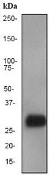
- Western blot - Prion protein PrP antibody [EP1802Y]; Anti-Prion protein PrP antibody [EP1802Y] at 1/10000 dilution + fetal brain lysate at 10 ug.Secondary.HRP-labelled goat anti-rabbit at 1/2000 dilution.Predicted band size : 28 kDa.
- Validation comment
- WB

- Submitted by
- OriGene (provider)
- Main image

- Experimental details
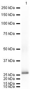
- Western blot ; Anti-Prion protein PrP antibody [EP1802Y] at 1/5000 dilution + Mouse Prion protein PrP full length protein at 0.01 ug.Secondary.Goat polyclonal Secondary Antibody to Rabbit IgG - H&L (HRP), pre-adsorbed at 1/5000 dilution.developed using the ECL technique.Performed under reducing conditions.Exposure time : 10 seconds
- Validation comment
- WB
Supportive validation
- Submitted by
- OriGene (provider)
- Main image

- Experimental details
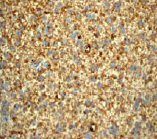
- Immunohistochemistry (Paraffin-embedded sections) - Prion protein PrP antibody [EP1802Y]; Immunohistochemical analysis of brain glioma using TA300958 at a dilution of 1/100-1/250.
- Validation comment
- IHC
- Submitted by
- OriGene (provider)
- Main image
- Experimental details
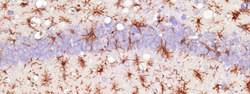
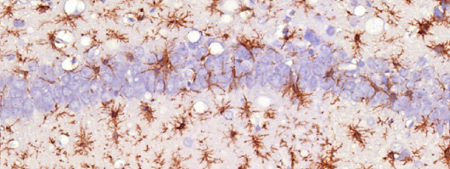
- Immunohistochemistry (Formalin/PFA-fixed paraffin-embedded sections) - Anti-Prion protein PrP antibody [EP1802Y]; Immunohistochemical analysis of Prion-infected mouse brain tissue, staining Prion protein PrP with TA300958. Antigen retrieval was performed by heat mediation in a citrate buffer (pH 6) before incubating with primary antibody (1/7000) overnight at 4?°C. Staining was detected using DAB.
- Validation comment
- IHC
- Submitted by
- OriGene (provider)
- Main image

- Experimental details
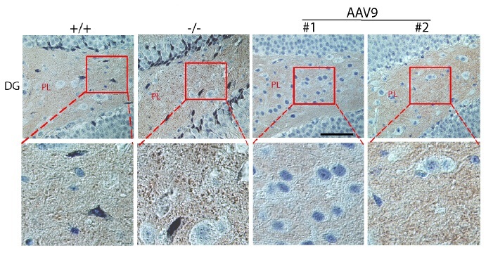
- Figure from citation: Immunohistochemistry of PRNP protein level by using anti-PRNP antibody in mouse brain tissue sections. Prnp-positive signals were stained brown. +/+: wt mice; -/-: non-treated MPS IIIB mice; AAV9: rAAV9-treated MPS IIIB mice; #1: mouse with low Prnp IHC intensity; #2: mouse with high Prnp IHC intensity; DG: dentate gyrus of hippocampus. PL: polymoph layer of DG. Scale bar: 50 um.
- Validation comment
- IHC
Supportive validation
- Submitted by
- OriGene (provider)
- Main image

- Experimental details
- Flow Cytometry-Anti-Prion protein PrP antibody(TA300958); Overlay histogram showing SH-SY5Y cells stained with TA300958 (red line). The secondary antibody used was DyLight 488 goat anti-rabbit IgG (H+L) at 1:500. Isotype control antibody (black line) was rabbit IgG (monoclonal) (1ug/1x10^6 cells) used under the same conditions. This antibody gave a positive signal in SH-SY5Y cells under the same conditions.
- Validation comment
- FC
