Explore
 Validate
 Learn
Learn Western blot
Antibody data
- Antibody Data
- Antigen structure
- References [1]
- Comments [0]
- Validations
- Western blot [5]
- Immunocytochemistry [1]
- Immunohistochemistry [1]
- Other assay [1]
Submit
Validation data
Reference
Comment
Report error
- Product number
- PA5-31293 - Provider product page

- Provider
- Invitrogen Antibodies
- Product name
- PDE10A Polyclonal Antibody
- Antibody type
- Polyclonal
- Antigen
- Recombinant protein fragment
- Reactivity
- Human, Mouse, Rat
- Host
- Rabbit
- Isotype
- IgG
- Vial size
- 100 µL
- Concentration
- 1.97 mg/mL
- Storage
- Store at 4°C short term. For long term storage, store at -20°C, avoiding freeze/thaw cycles.
Submitted references Phosphodiesterase 10A IgG: A novel biomarker of paraneoplastic neurologic autoimmunity.
Zekeridou A, Kryzer T, Guo Y, Hassan A, Lennon V, Lucchinetti CF, Pittock S, McKeon A
Neurology 2019 Aug 20;93(8):e815-e822
Neurology 2019 Aug 20;93(8):e815-e822
No comments: Submit comment
Supportive validation
- Submitted by
- Invitrogen Antibodies (provider)
- Main image

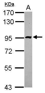
- Experimental details
- Western blot analysis of PDE10A using 30 µg of HeLa lysate. Samples were loaded onto a 7.5% SDS-PAGE gel and probed with a PDE10A polyclonal antibody (Product # PA5-31293) at a dilution of 1:3000.
- Submitted by
- Invitrogen Antibodies (provider)
- Main image
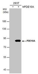
- Experimental details
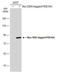
- Western blot analysis of PDE10A in Non-transfected (-) and transfected (+) 293T whole cell extracts (30 µg). Samples were separated by 7.5% SDS-PAGE and the membrane was probed with PDE10A Polyclonal antibody (Product # PA5-31293) at a dilution of 1:5000.
- Submitted by
- Invitrogen Antibodies (provider)
- Main image
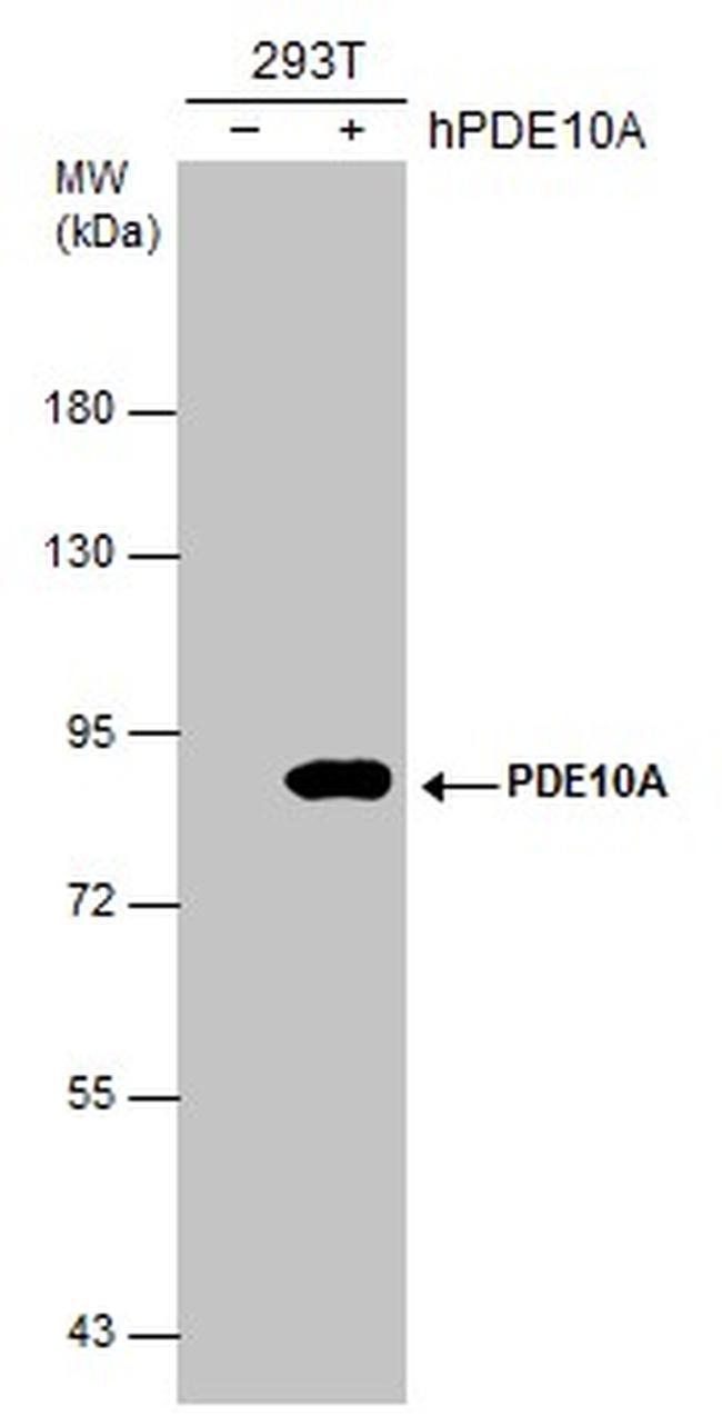
- Experimental details
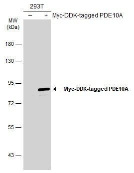
- Western Blot analysis of PDE10A was performed by separating 30 µg of non-transfected (–) and transfected (+) 293T whole cell extracts by 5% SDS-PAGE. Proteins were transferred to a membrane and probed with a PDE10A Polyclonal Antibody (Product # PA5-31293) at a dilution of 1:10000. The HRP-conjugated anti-rabbit IgG antibody was used to detect the primary antibody.
- Submitted by
- Invitrogen Antibodies (provider)
- Main image
- Experimental details
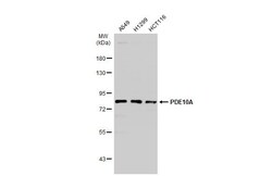
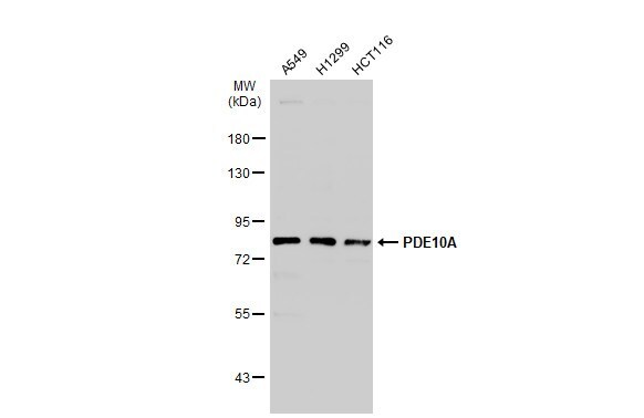
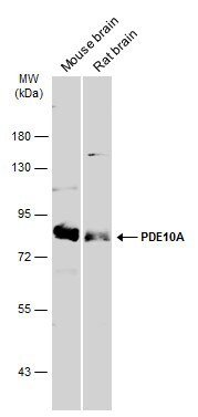
- Western Blot using PDE10A Polyclonal Antibody (Product # PA5-31293). Various whole cell extracts (30 µg) were separated by 7.5% SDS-PAGE, and the membrane was blotted with PDE10A Polyclonal Antibody (Product # PA5-31293) diluted at 1:1,000. The HRP-conjugated anti-rabbit IgG antibody was used to detect the primary antibody.
- Submitted by
- Invitrogen Antibodies (provider)
- Main image
- Experimental details
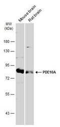
- Western Blot using PDE10A Polyclonal Antibody (Product # PA5-31293). Various tissue extracts (50 µg) were separated by 7.5% SDS-PAGE, and the membrane was blotted with PDE10A Polyclonal Antibody (Product # PA5-31293) diluted at 1:3,000. The HRP-conjugated anti-rabbit IgG antibody was used to detect the primary antibody.
Supportive validation
- Submitted by
- Invitrogen Antibodies (provider)
- Main image

- Experimental details
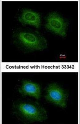
- Immunofluorescent analysis of PDE10A in methanol-fixed HeLa cells using a PDE10A polyclonal antibody (Product # PA5-31293) at a 1:200 dilution.
Supportive validation
- Submitted by
- Invitrogen Antibodies (provider)
- Main image
- Experimental details
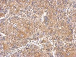
- Immunohistochemical analysis of paraffin-embedded Cal27 xenograft, using PDE10A (Product # PA5-31293) antibody at 1:500 dilution. Antigen Retrieval: EDTA based buffer, pH 8.0, 15 min.

Supportive validation
- Submitted by
- Invitrogen Antibodies (provider)
- Main image
- Experimental details
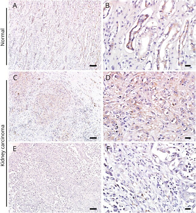
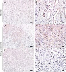
- Figure 3 Immunohistochemistry of renal cell carcinoma (patient 4) (A, B) Phosphodiesterase 10A (PDE10A) immunohistochemistry in normal control human kidney tissue. The high power image (B) indicates the collecting tubular epithelium with marked expression of PDE10A. The kidney carcinoma tissue of patient 4 shows foci of variable PDE10A immunoreactivity: moderate expression (C, D) or scant PDE10A expression (E, F) in different parts of the tumor. Scale bars in A, C, and E = 100 mum. Scale bars in B, D, and F = 20 mum.