Explore
 Validate
 Learn
Learn Western blot
Antibody data
- Antibody Data
- Antigen structure
- References [3]
- Comments [0]
- Validations
- Western blot [1]
- Flow cytometry [1]
Submit
Validation data
Reference
Comment
Report error
- Product number
- PA3-112 - Provider product page

- Provider
- Invitrogen Antibodies
- Product name
- SSTR5 Polyclonal Antibody
- Antibody type
- Polyclonal
- Antigen
- Synthetic peptide
- Description
- PA3-112 detects the Somatostatin Receptor 5 in human samples.
- Concentration
- Conc. Not Determined
Submitted references Pituispheres Contain Genetic Variants Characteristic to Pituitary Adenoma Tumor Tissue.
Combined effects of octreotide and cisplatin on the proliferation of side population cells from anaplastic thyroid cancer cell lines.
Somatostatin receptor subtypes in human pheochromocytoma: subcellular expression pattern and functional relevance for octreotide scintigraphy.
Peculis R, Mandrika I, Petrovska R, Dortane R, Megnis K, Nazarovs J, Balcere I, Stukens J, Konrade I, Pirags V, Klovins J, Rovite V
Frontiers in endocrinology 2020;11:313
Frontiers in endocrinology 2020;11:313
Combined effects of octreotide and cisplatin on the proliferation of side population cells from anaplastic thyroid cancer cell lines.
Li Z, Jiang X, Chen P, Wu X, Duan A, Qin Y
Oncology letters 2018 Sep;16(3):4033-4042
Oncology letters 2018 Sep;16(3):4033-4042
Somatostatin receptor subtypes in human pheochromocytoma: subcellular expression pattern and functional relevance for octreotide scintigraphy.
Mundschenk J, Unger N, Schulz S, Höllt V, Schulz S, Steinke R, Lehnert H
The Journal of clinical endocrinology and metabolism 2003 Nov;88(11):5150-7
The Journal of clinical endocrinology and metabolism 2003 Nov;88(11):5150-7
No comments: Submit comment
Supportive validation
- Submitted by
- Invitrogen Antibodies (provider)
- Main image

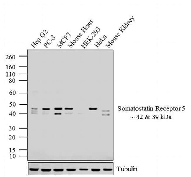
- Experimental details
- Western blot analysis was performed on whole cell extracts (20 µg lysate) of Hep G2 (Lane 1), PC-3 (Lane 2), MCF7 (Lane 3), Mouse Heart (lane 4), HEK-293 (lane 5), HeLa (lane 6) and Mouse Kidney (lane 9). The blots were probed with Anti-Somatostatin Receptor 5 Rabbit Polyclonal Antibody (Product # PA3-112, 1:4000-1:6000 dilution) and detected by chemiluminescence using Goat anti-Rabbit IgG (H+L) Superclonal™ Secondary Antibody, HRP conjugate (Product # A27036, 0.4 µg/mL, 1:2500 dilution). Two bands ~ 42 and 39 kDa corresponding to Somatostatin Receptor 5 was observed across cell lines and tissues tested except for HEK-293. Known quantity of protein samples were electrophoresed using Novex® NuPAGE® 4-12 % Bis-Tris gel (Product # NP0322BOX), XCell SureLock™ Electrophoresis System (Product # EI0002) and Novex® Sharp Pre-Stained Protein Standard (Product # LC5800). Resolved proteins were then transferred onto a nitrocellulose membrane with iBlot® 2 Dry Blotting System (Product # IB21001). The membrane was probed with the relevant primary and secondary Antibody using iBind™ Flex Western Starter Kit (Product # SLF2000S). Chemiluminescent detection was performed using Pierce™ ECL Western Blotting Substrate (Product # 32106).
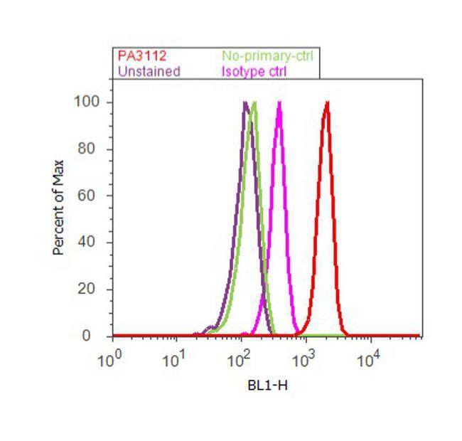
Supportive validation
- Submitted by
- Invitrogen Antibodies (provider)
- Main image

- Experimental details
- Flow cytometry analysis of Somatostatin Receptor 5 was done on HT-29 cells. Cells were fixed with 70% ethanol for 10 minutes, permeabilized with 0.25% Triton™ X-100 for 20 minutes, and blocked with 5% BSA for 30 minutes at room temperature. Cells were labeled with Somatostatin Receptor 5 Rabbit Polyclonal Antibody (PA3112, red histogram) or with rabbit isotype control (pink histogram) at 3-5 ug/million cells in 2.5% BSA. After incubation at room temperature for 2 hours, the cells were labeled with Alexa Fluor® 488 Goat Anti-Rabbit Secondary Antibody (A11008) at a dilution of 1:400 for 30 minutes at room temperature. The representative 10,000 cells were acquired and analyzed for each sample using an Attune® Acoustic Focusing Cytometer. The purple histogram represents unstained control cells and the green histogram represents no-primary-antibody control.