 Explore
 Validate
 Learn
Learn Western blot
Antibody data
- Antibody Data
- Antigen structure
- References [1]
- Comments [0]
- Validations
- Western blot [7]
- Immunocytochemistry [1]
- Immunohistochemistry [2]
Submit
Validation data
Reference
Comment
Report error
- Product number
- PA5-27539 - Provider product page

- Provider
- Invitrogen Antibodies
- Product name
- RPL5 Polyclonal Antibody
- Antibody type
- Polyclonal
- Antigen
- Recombinant protein fragment
- Description
- Recommended positive controls: 293T, A431, HeLa, HepG2, Mouse brain, Rat2.
- Concentration
- 0.4 mg/mL
Submitted references A single synonymous mutation determines the phosphorylation and stability of the nascent protein.
Karakostis K, Vadivel Gnanasundram S, López I, Thermou A, Wang L, Nylander K, Olivares-Illana V, Fåhraeus R
Journal of molecular cell biology 2019 Mar 1;11(3):187-199
Journal of molecular cell biology 2019 Mar 1;11(3):187-199
No comments: Submit comment
Supportive validation
- Submitted by
- Invitrogen Antibodies (provider)
- Main image

- Experimental details
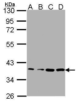
- Western blot analysis of RPL5 using 30µg of A) A431 (B) H1299 (C) HeLaS3 and D) MOLT4 lysate. Samples were loaded onto a 12% SDS-PAGE gel and probed with a RPL5 polyclonal antibody (Product # PA5-27539) at a dilution of 1:1000.
- Submitted by
- Invitrogen Antibodies (provider)
- Main image
- Experimental details
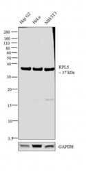
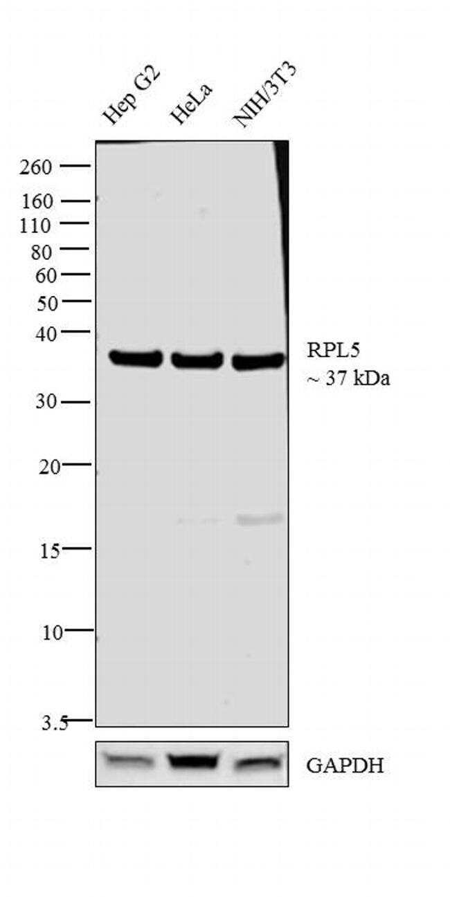
- Western blot analysis was performed on whole cell extracts (30 µg lysate) of Hep G2 (Lane 1), HeLa (Lane 2) and NIH/3T3 (Lane 3). The blot was probed with Anti-RPL5 Polyclonal Antibody (Product # PA5-27539, 1:1000 dilution) and detected by chemiluminescence using Goat anti-Rabbit IgG (H+L) Superclonal™ Secondary Antibody, HRP conjugate (Product # A27036, 0.25 µg/ml, 1:4000 dilution). A 37 kDa band corresponding to RPL5 was observed across the cell lines tested.
- Submitted by
- Invitrogen Antibodies (provider)
- Main image
- Experimental details
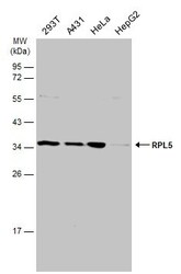
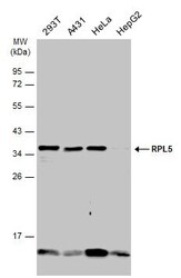
- Western Blot analysis of RPL5 was performed by separating 30 µg of various whole cell extracts by 12% SDS-PAGE. Proteins were transferred to a membrane and probed with a RPL5 Polyclonal Antibody (Product # PA5-27539) at a dilution of 1:1000 and a HRP-conjugated anti-rabbit IgG secondary antibody.
- Submitted by
- Invitrogen Antibodies (provider)
- Main image

- Experimental details
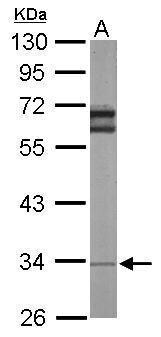
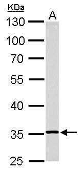
- Western Blot using RPL5 Polyclonal Antibody (Product # PA5-27539). Sample (50 µg of whole cell lysate). Lane A: Mouse brain. 10% SDS PAGE. RPL5 Polyclonal Antibody (Product # PA5-27539) diluted at 1:1,000. The HRP-conjugated anti-rabbit IgG antibody was used to detect the primary antibody.
- Submitted by
- Invitrogen Antibodies (provider)
- Main image
- Experimental details
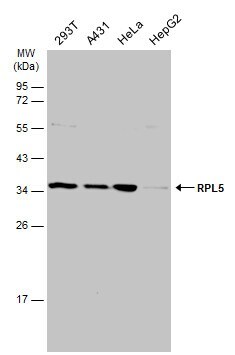
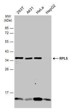
- Western Blot using RPL5 Polyclonal Antibody (Product # PA5-27539). Various whole cell extracts (30 µg) were separated by 12% SDS-PAGE, and the membrane was blotted with RPL5 Polyclonal Antibody (Product # PA5-27539) diluted at 1:1,000. The HRP-conjugated anti-rabbit IgG antibody was used to detect the primary antibody.
- Submitted by
- Invitrogen Antibodies (provider)
- Main image

- Experimental details
- RPL5 Polyclonal Antibody detects RPL5 protein by western blot analysis. A. 30 µg Rat2 whole cell lysate/extract.10% SDS-PAGE. RPL5 Polyclonal Antibody (Product # PA5-27539) dilution: 1:1,000. The HRP-conjugated anti-rabbit IgG antibody was used to detect the primary antibody.
- Submitted by
- Invitrogen Antibodies (provider)
- Main image
- Experimental details
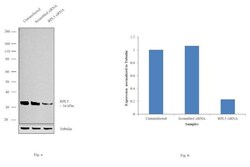
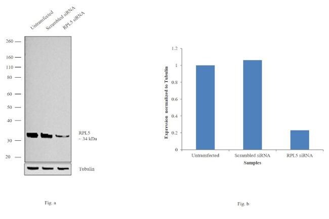
- Knockdown of RPL5 was achieved by transfecting HeLa cells with RPL5 specific siRNAs (Silencer® select Product # s12152). Western blot analysis (Fig. a) was performed using whole cell extracts from the RPL5 knockdown cells (lane 3), non-specific scrambled siRNA transfected cells (lane 2) and untransfected cells (lane 1). The blot was probed with RPL5 Polyclonal Antibody (Product # PA5-27539, 1:2000 dilution) and Goat anti-Rabbit IgG (H+L) Superclonal™ Secondary Antibody, HRP conjugate (Product # A27036, 0.25µg/ml, 1:4000 dilution). Densitometric analysis of this western blot is shown in histogram (Fig. b). Decrease in signal upon siRNA mediated knock down confirms that antibody is specific to RPL5.
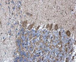
Supportive validation
- Submitted by
- Invitrogen Antibodies (provider)
- Main image

- Experimental details
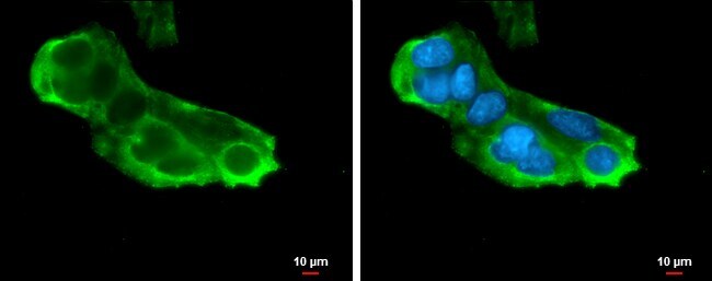
- Immunocytochemistry-Immunofluorescence analysis of RPL5 was performed in HepG2 cells fixed in 4% paraformaldehyde at RT for 15 min. Green: RPL5 Polyclonal Antibody (Product # PA5-27539) diluted at 1:500. Blue: Hoechst 33342 staining. Scale bar = 10 µm.
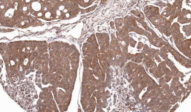
Supportive validation
- Submitted by
- Invitrogen Antibodies (provider)
- Main image
- Experimental details
- Immunohistochemical analysis of paraffin-embedded Gastric CA N87 xenograft, using RPL5 (Product # PA5-27539) antibody at 1:100 dilution. Antigen Retrieval: EDTA based buffer, pH 8.0, 15 min.
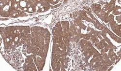
- Submitted by
- Invitrogen Antibodies (provider)
- Main image
- Experimental details
- RPL5 Polyclonal Antibody detects RPL5 protein at cytoplasm on mouse hind brain by immunohistochemical analysis. Sample: Paraffin-embedded mouse hind brain. RPL5 Polyclonal Antibody (Product # PA5-27539) diluted at 1:500. Antigen Retrieval: EDTA based buffer, pH 8.0, 15 min.
