Antibody data
- Antibody Data
- Antigen structure
- References [1]
- Comments [0]
- Validations
- Western blot [4]
- Immunocytochemistry [2]
- Immunohistochemistry [1]
- Other assay [2]
Submit
Validation data
Reference
Comment
Report error
- Product number
- PA5-95826 - Provider product page

- Provider
- Invitrogen Antibodies
- Product name
- PYCARD Polyclonal Antibody
- Antibody type
- Polyclonal
- Antigen
- Recombinant full-length protein
- Description
- Positive Samples: HT-29, BT-474, U-937, mouse thymus, mouse spleen, rat spleen; Cellular Location: Cytoplasm, Endoplasmic reticulum, Mitochondrion, Nucleus Immunogen sequence: MGRARDAILD ALENLTAEEL KKFKLKLLSV PLREGYGRIP RGALLSMDAL DLTDKLVSFY LETYGAELTA NVLRDMGLQE MAGQLQAATH Q
- Reactivity
- Human, Mouse, Rat
- Host
- Rabbit
- Isotype
- IgG
- Vial size
- 100 µL
- Concentration
- 2.65 mg/mL
- Storage
- -20° C, Avoid Freeze/Thaw Cycles
Submitted references Colchicine Ameliorates Dilated Cardiomyopathy Via SIRT2-Mediated Suppression of NLRP3 Inflammasome Activation.
Sun X, Duan J, Gong C, Feng Y, Hu J, Gu R, Xu B
Journal of the American Heart Association 2022 Jul 5;11(13):e025266
Journal of the American Heart Association 2022 Jul 5;11(13):e025266
No comments: Submit comment
Supportive validation
- Submitted by
- Invitrogen Antibodies (provider)
- Main image

- Experimental details
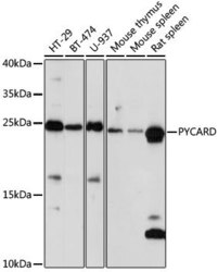
- Western blot analysis of extracts of various cell lines, using PYCARD Polyclonal antibody (Product # PA5-95826) at 1:1000 dilution. Secondary antibody: HRP Goat Anti-Rabbit IgG (H+L) at 1:10000 dilution. Lysates/proteins: 25ug per lane. Blocking buffer: 3% nonfat dry milk in TBST. Exposure time: 30s.
- Submitted by
- Invitrogen Antibodies (provider)
- Main image

- Experimental details
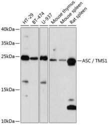
- Western Blot analysis of PYCARD in extracts of various cell lines using PYCARD Polyclonal Antibody (Product # PA5-95826) at a dilution of 1:1000. A HRP Goat Anti-Rabbit IgG (H+L) secondary antibody was used at a dilution of 1:10,000. Lysates/proteins: 25 µg per lane. Blocking buffer: 3% nonfat dry milk in TBST.
- Submitted by
- Invitrogen Antibodies (provider)
- Main image

- Experimental details
- Knockout of PYCARD was achieved by CRISPR-Cas9 genome editing using LentiArray™ Lentiviral sgRNA (Product # A32042, Assay ID CRISPR1008587_LV) and LentiArray Cas9 Lentivirus (Product # A32064). Western blot analysis of PYCARD was performed by loading 20 µg of THP-1 wild type (Lane-1), THP-1 Cas9 (Lane-2) and THP-1 PYCARD KO (Lane-3) whole cell extracts. The samples were electrophoresed using NuPAGE™ Novex™ 4-12% Bis-Tris Protein Gel (Product # NP0322BOX). Resolved proteins were then transferred onto a nitrocellulose membrane (Product # IB23001) by iBlot® 2 Dry Blotting System (Product # IB21001). The blot was probed with PYCARD Polyclonal Antibody (Product # PA5-95826, 1:2000 dilution) and Goat anti-Rabbit IgG (H+L) Superclonal™ Recombinant Secondary Antibody, HRP (Product # A27036, 1:10,000 dilution) using the iBright™ FL1500 (Product # A44115). Chemiluminescent detection was performed using Novex® ECL Chemiluminescent Substrate Reagent Kit (Product # WP20005). Loss of signal upon CRISPR mediated knockout (KO) using the LentiArray™ CRISPR product line confirms that antibody is specific to PYCARD. Uncharacterized band was observed in ~100 kDa in all samples.
- Submitted by
- Invitrogen Antibodies (provider)
- Main image

- Experimental details
- Western blot was performed using Anti-PYCARD Polyclonal Antibody (Product # PA5-95826) and a 21kDa band corresponding to Apoptosis-associated speck-like protein containing a CARD was observed across the cell lines tested and tissues tested. Whole cell extracts (30 µg lysate) of MCF7 (Lane 1), THP-1 (Lane 2), HL-60 (Lane 3), K-562 (Lane 4), HEK-293 (Lane 5), HeLa (Lane 6), Jurkat (Lane 7), Mouse Spleen (Lane 8), Rat Spleen (Lane 9), Mouse Liver (Lane 10), Rat Liver (Lane 11) were electrophoresed using NuPAGE™ 4-12% Bis-Tris Protein Gel (Product # NP0322BOX). Resolved proteins were then transferred onto a Nitrocellulose membrane (Product # IB23002) by iBlot® 2 Dry Blotting System (Product # IB21001). The blot was probed with the primary antibody (1:100) and detected by chemiluminescence with Goat anti-Rabbit IgG (H+L) Superclonal™ Recombinant Secondary Antibody, HRP (Product # A27036, 1:4000) using the iBright FL 1000 (Product # A32752). Chemiluminescent detection was performed using Novex® ECL Chemiluminescent Substrate Reagent Kit (Product # WP20005).
Supportive validation
- Submitted by
- Invitrogen Antibodies (provider)
- Main image

- Experimental details
- Immunocytochemistry-Immunofluorescence analysis of PYCARD was performed in U-2 OS cells using PYCARD Polyclonal Antibody (Product # PA5-95826).
- Submitted by
- Invitrogen Antibodies (provider)
- Main image

- Experimental details
- Immunocytochemistry-Immunofluorescence analysis of PYCARD was performed in U-2 OS cells using PYCARD Polyclonal Antibody (Product # PA5-95826).
Supportive validation
- Submitted by
- Invitrogen Antibodies (provider)
- Main image

- Experimental details
- Immunohistochemistry analysis of PYCARD in paraffin-embedded human placenta using PYCARD Polyclonal Antibody (Product # PA5-95826) at a dilution of 1:200.
Supportive validation
- Submitted by
- Invitrogen Antibodies (provider)
- Main image

- Experimental details
- Immunoprecipitation analysis of PYCARD was performed in 200 µg extracts of U937 cells using PYCARD Polyclonal Antibody (Product # PA5-95826). Western blot was performed from the immunoprecipitate using PYCARD Polyclonal Antibody.
- Submitted by
- Invitrogen Antibodies (provider)
- Main image

- Experimental details
- Figure 3 NLRP3 (NOD-like receptor protein 3) inflammasome pathway was inhibited by colchicine treatment. A and B , Representative immunoblots and the corresponding analysis of NLRP3 inflammasome pathway in cardiac tissues at the end point of experiment. GAPDH shows as loading control. C , Representative immunofluorescence images of cardiac tissues staining with ASC (green), cTNT (cardiac troponin T) (red), and DAPI (blue). Frequency of ASC speck is shown on the right (n=6-8 each group). Five images per mouse are evaluated. Scale bar=50 mum. Data are shown as mean+-SEM. All data statistical significance are determined by 1-way ANOVA (Tukey post-test). cTNT indicates cardiac troponin T; DCM, dilated cardiomyopathy; IL-1beta, interleukin-1beta; and NLRP3, NOD-like receptor protein 3. * P
 Explore
 Validate
 Learn
Learn Western blot
 Immunoprecipitation