Explore
 Validate
 Learn
Learn Western blot
 Immunoprecipitation
Antibody data
- Antibody Data
- Antigen structure
- References [8]
- Comments [0]
- Validations
- Western blot [1]
- Immunohistochemistry [2]
Submit
Validation data
Reference
Comment
Report error
- Product number
- AF3715 - Provider product page

- Provider
- R&D Systems
- Product name
- Human Fibroblast Activation Protein alpha/FAP Antibody
- Antibody type
- Polyclonal
- Description
- Immunogen affinity purified. Detects human Fibroblast Activation Protein alpha/FAP in direct ELISAs and Western blots.
- Reactivity
- Human
- Host
- Sheep
- Conjugate
- Unconjugated
- Antigen sequence
Q12884- Isotype
- IgG
- Vial size
- 100 ug
- Concentration
- LYOPH
- Storage
- Use a manual defrost freezer and avoid repeated freeze-thaw cycles. 12 months from date of receipt, -20 to -70 °C as supplied. 1 month, 2 to 8 °C under sterile conditions after reconstitution. 6 months, -20 to -70 °C under sterile conditions after reconstitution.
Submitted references Fibroblast activation protein-positive fibroblasts promote tumor progression through secretion of CCL2 and interleukin-6 in esophageal squamous cell carcinoma.
Tumor-targeting efficacy of a BF211 prodrug through hydrolysis by fibroblast activation protein-α.
Cancer-associated fibroblasts induce antigen-specific deletion of CD8 (+) T Cells to protect tumour cells.
A synthetic urinary probe-coated nanoparticles sensitive to fibroblast activation protein α for solid tumor diagnosis.
Fibroblast activation protein augments progression and metastasis of pancreatic ductal adenocarcinoma.
Adrenergic-mediated increases in INHBA drive CAF phenotype and collagens.
Fibroblast Activation Protein (FAP) Accelerates Collagen Degradation and Clearance from Lungs in Mice.
Antitumor effects of chimeric receptor engineered human T cells directed to tumor stroma.
Higashino N, Koma YI, Hosono M, Takase N, Okamoto M, Kodaira H, Nishio M, Shigeoka M, Kakeji Y, Yokozaki H
Laboratory investigation; a journal of technical methods and pathology 2019 Jun;99(6):777-792
Laboratory investigation; a journal of technical methods and pathology 2019 Jun;99(6):777-792
Tumor-targeting efficacy of a BF211 prodrug through hydrolysis by fibroblast activation protein-α.
Chai XP, Sun GL, Fang YF, Hu LH, Liu X, Zhang XW
Acta pharmacologica Sinica 2018 Mar;39(3):415-424
Acta pharmacologica Sinica 2018 Mar;39(3):415-424
Cancer-associated fibroblasts induce antigen-specific deletion of CD8 (+) T Cells to protect tumour cells.
Lakins MA, Ghorani E, Munir H, Martins CP, Shields JD
Nature communications 2018 Mar 5;9(1):948
Nature communications 2018 Mar 5;9(1):948
A synthetic urinary probe-coated nanoparticles sensitive to fibroblast activation protein α for solid tumor diagnosis.
Feng X, Wang Q, Liao Y, Zhou X, Wang Y, Liu W, Zhang G
International journal of nanomedicine 2017;12:5359-5372
International journal of nanomedicine 2017;12:5359-5372
Fibroblast activation protein augments progression and metastasis of pancreatic ductal adenocarcinoma.
Lo A, Li CP, Buza EL, Blomberg R, Govindaraju P, Avery D, Monslow J, Hsiao M, Puré E
JCI insight 2017 Oct 5;2(19)
JCI insight 2017 Oct 5;2(19)
Adrenergic-mediated increases in INHBA drive CAF phenotype and collagens.
Nagaraja AS, Dood RL, Armaiz-Pena G, Kang Y, Wu SY, Allen JK, Jennings NB, Mangala LS, Pradeep S, Lyons Y, Haemmerle M, Gharpure KM, Sadaoui NC, Rodriguez-Aguayo C, Ivan C, Wang Y, Baggerly K, Ram P, Lopez-Berestein G, Liu J, Mok SC, Cohen L, Lutgendorf SK, Cole SW, Sood AK
JCI insight 2017 Aug 17;2(16)
JCI insight 2017 Aug 17;2(16)
Fibroblast Activation Protein (FAP) Accelerates Collagen Degradation and Clearance from Lungs in Mice.
Fan MH, Zhu Q, Li HH, Ra HJ, Majumdar S, Gulick DL, Jerome JA, Madsen DH, Christofidou-Solomidou M, Speicher DW, Bachovchin WW, Feghali-Bostwick C, Puré E
The Journal of biological chemistry 2016 Apr 8;291(15):8070-89
The Journal of biological chemistry 2016 Apr 8;291(15):8070-89
Antitumor effects of chimeric receptor engineered human T cells directed to tumor stroma.
Kakarla S, Chow KK, Mata M, Shaffer DR, Song XT, Wu MF, Liu H, Wang LL, Rowley DR, Pfizenmaier K, Gottschalk S
Molecular therapy : the journal of the American Society of Gene Therapy 2013 Aug;21(8):1611-20
Molecular therapy : the journal of the American Society of Gene Therapy 2013 Aug;21(8):1611-20
No comments: Submit comment
Supportive validation
- Submitted by
- R&D Systems (provider)
- Main image

- Experimental details
- Detection of Human Fibroblast Activation Protein alpha/FAP by Western Blot. Western blot shows lysates of WI-38 human lung fibroblast cell line. PVDF membrane was probed with 0.5 µg/mL of Sheep Anti-Human Fibroblast Activation Protein alpha/FAP Antigen Affinity-purified Polyclonal Antibody (Catalog # AF3715) followed by HRP-conjugated Anti-Sheep IgG Secondary Antibody (Catalog # HAF016). A specific band was detected for Fibroblast Activation Protein alpha/FAP at approximately 97 kDa (as indicated). This experiment was conducted under reducing conditions and using Immunoblot Buffer Group 1.

Supportive validation
- Submitted by
- R&D Systems (provider)
- Main image

- Experimental details
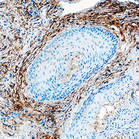
- Fibroblast Activation Protein alpha/FAP in Human Squamous Cell Carcinoma. Fibroblast Activation Protein alpha/FAP was detected in immersion fixed paraffin-embedded sections of human squamous cell carcinoma using Sheep Anti-Human Fibroblast Activation Protein alpha/FAP Antigen Affinity-purified Polyclonal Antibody (Catalog # AF3715) at 15 µg/mL overnight at 4 °C. Tissue was stained using the Anti-Sheep HRP-DAB Cell & Tissue Staining Kit (brown; Catalog # CTS019) and counterstained with hematoxylin (blue). Specific staining was localized to connective tissue. View our protocol for Chromogenic IHC Staining of Paraffin-embedded Tissue Sections.
- Submitted by
- R&D Systems (provider)
- Main image

- Experimental details
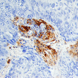
- Fibroblast Activation Protein alpha/FAP in Human Basal Cell Carcinoma. Fibroblast Activation Protein alpha/FAP was detected in immersion fixed paraffin-embedded sections of human basal cell carcinoma using Sheep Anti-Human Fibroblast Activation Protein alpha/FAP Antigen Affinity-purified Polyclonal Antibody (Catalog # AF3715) at 10 µg/mL overnight at 4 °C. Tissue was stained using the Anti-Sheep HRP-DAB Cell & Tissue Staining Kit (brown; Catalog # CTS019) and counterstained with hematoxylin (blue). Specific staining was localized to cytoplasm. View our protocol for Chromogenic IHC Staining of Paraffin-embedded Tissue Sections.