Explore
 Validate
 Learn
Learn Western blot
Antibody data
- Antibody Data
- Antigen structure
- References [2]
- Comments [0]
- Validations
- Western blot [2]
- Immunocytochemistry [4]
- Flow cytometry [1]
- Other assay [1]
Submit
Validation data
Reference
Comment
Report error
- Product number
- PA1-007 - Provider product page

- Provider
- Invitrogen Antibodies
- Product name
- ERp72 Polyclonal Antibody
- Antibody type
- Polyclonal
- Antigen
- Synthetic peptide
- Description
- PA1-007 detects ERp72 protein in human and mouse samples. PA1-007 has successfully been used in ICC, IF and Western blot procedures. By Western blot, this antibody detects a 72 kDa protein representing ERp72 in human fibrosarcoma HT 1080 cell lysate. The PA1-007 immunogen is a synthetic peptide corresponding to residues K(629) F I E E H A T K L S R T K E E L(645) of humna ERp72. This peptide (Cat. # PEP-223) is available for use in neutralization and control experiments.
- Reactivity
- Human, Mouse
- Host
- Rabbit
- Isotype
- IgG
- Vial size
- 100 µg
- Concentration
- 1 mg/mL
- Storage
- -20° C, Avoid Freeze/Thaw Cycles
Submitted references Protein Disulfide Isomerase A4 Is Involved in Genome Uncoating during Human Astrovirus Cell Entry.
Axons provide the secretory machinery for trafficking of voltage-gated sodium channels in peripheral nerve.
Aguilar-Hernández N, Meyer L, López S, DuBois RM, Arias CF
Viruses 2020 Dec 31;13(1)
Viruses 2020 Dec 31;13(1)
Axons provide the secretory machinery for trafficking of voltage-gated sodium channels in peripheral nerve.
González C, Cánovas J, Fresno J, Couve E, Court FA, Couve A
Proceedings of the National Academy of Sciences of the United States of America 2016 Feb 16;113(7):1823-8
Proceedings of the National Academy of Sciences of the United States of America 2016 Feb 16;113(7):1823-8
No comments: Submit comment
Supportive validation
- Submitted by
- Invitrogen Antibodies (provider)
- Main image
- Experimental details
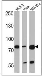
- Western blot analysis of ERp72 was performed by loading 25 µg of MCF-7 (lane 1), Hela (lane 2) and NIH-3T3 (lane 3) lysates onto an SDS polyacrylamide gel. Proteins were transferred to a PVDF membrane and blocked at 4ºC overnight. The membrane was probed with an ERp72 polyclonal antibody (Product # PA1-007) at a dilution of 1:1000 overnight at 4°C, washed in TBST, and probed with an HRP-conjugated goat anti-rabbit IgG (H+L) cross-adsorbed secondary antibody for 1 hr at room temperature in the dark. Chemiluminescent detection was performed using Pierce ECL Plus Western Blotting Substrate (Product # 32132). Results show a band at ~72 kDa.

- Submitted by
- Invitrogen Antibodies (provider)
- Main image
- Experimental details
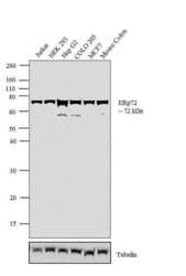
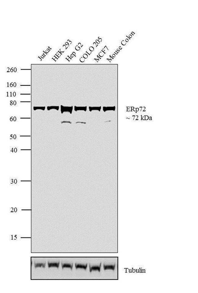
- Western blot analysis was performed on whole cell extracts (30 µg lysate) of Jurkat (Lane 1), HEK 293 (Lane 2), Hep G2 (Lane 3), COLO 205 (Lane 4), MCF7 (Lane 5) and tissue extract of Mouse Colon (Lane 6). The blots were probed with Anti-ERp72 Rabbit Polyclonal Antibody (Product # PA1-007, 1:100-1:1000) and detected by chemiluminescence using Goat anti-Rabbit IgG (H+L) Superclonal™ Secondary Antibody, HRP conjugate (Product # A27036, 0.4 µg/mL, 1:2500 dilution). A 72 kDa band corresponding to ERp72 was observed across the cell lines and tissue tested. Known quantity of protein samples were electrophoresed using Novex® NuPAGE® 10 % Bis-Tris gel (Product # NP0301BOX), XCell SureLock™ Electrophoresis System (Product # EI0002) and Novex® Sharp Pre-Stained Protein Standard (Product # LC5800). Resolved proteins were then transferred onto a nitrocellulose membrane with iBlot® 2 Dry Blotting System (Product # IB21001). The membrane was probed with the relevant primary and secondary Antibody using iBind™ Flex Western Starter Kit (Product # SLF2000S). Chemiluminescent detection was performed using Pierce™ ECL Western Blotting Substrate (Product # 32106).
Supportive validation
- Submitted by
- Invitrogen Antibodies (provider)
- Main image
- Experimental details
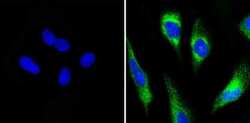
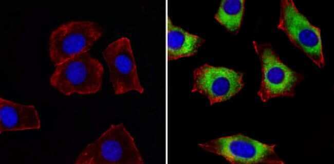
- Immunofluorescent analysis of ERp72 (green) showing ER staining in Hela cells (right) compared to a negative control without primary antibody (left). Cells were permeabilized with 0.1% Triton X-100 in TBS for 5-10 minutes and blocked with 3% BSA-PBS for 30 minutes at room temperature. Cells were probed with a ERp72 polyclonal antibody (Product # PA1-007) in 3% BSA-PBS at a dilution of 1:100 and incubated overnight at 4ºC in a humidified chamber. Cells were washed with PBST and incubated with a DyLight-488 conjugated secondary antibody in PBS at room temperature in the dark. Nuclei were stained with DAPI (blue) and images were taken at a magnification of 60x.
- Submitted by
- Invitrogen Antibodies (provider)
- Main image
- Experimental details
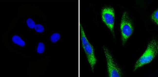
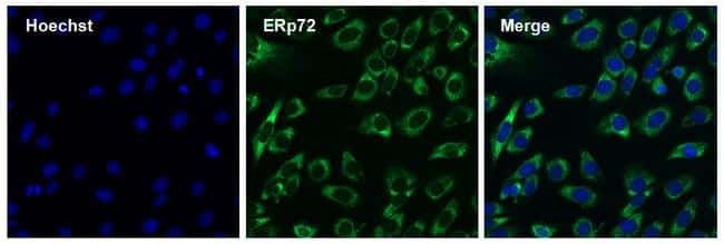
- Immunofluorescent analysis of ERp72 (green) showing ER staining in MCF-7 cells (right) compared to a negative control without primary antibody (left). Cells were permeabilized with 0.1% Triton X-100 in TBS for 5-10 minutes and blocked with 3% BSA-PBS for 30 minutes at room temperature. Cells were probed with a ERp72 polyclonal antibody (Product # PA1-007) in 3% BSA-PBS at a dilution of 1:100 and incubated overnight at 4ºC in a humidified chamber. Cells were washed with PBST and incubated with a DyLight-488 conjugated secondary antibody in PBS at room temperature in the dark. Nuclei were stained with DAPI (blue) and images were taken at a magnification of 60x.

- Submitted by
- Invitrogen Antibodies (provider)
- Main image
- Experimental details
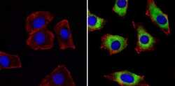
- Immunofluorescent analysis of ERp72 (green) showing ER staining in NIH-3T3 cells (right) compared to a negative control without primary antibody (left). Cells were permeabilized with 0.1% Triton X-100 in TBS for 5-10 minutes and blocked with 3% BSA-PBS for 30 minutes at room temperature. Cells were probed with a ERp72 polyclonal antibody (Product # PA1-007) in 3% BSA-PBS at a dilution of 1:100 and incubated overnight at 4ºC in a humidified chamber. Cells were washed with PBST and incubated with a DyLight-488 conjugated secondary antibody in PBS at room temperature in the dark. Actin was stained with a Dylight 554 phalloidin (red) and nuclei were stained with DAPI (blue). Images were taken at a magnification of 60x.
- Submitted by
- Invitrogen Antibodies (provider)
- Main image
- Experimental details
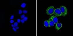
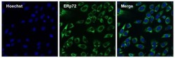
- Immunofluorescent analysis of ERp72 (green) in 3T3 cells. The cells were permeabilized with 0.1% Triton X-100 in TBS for 15 minutes, and blocked with 3% Blocker BSA in PBS (Product # 37525) for 15 minutes at room temperature. Cells were stained with a ERp72 rabbit polyclonal antibody (Product # PA1-007), at a concentration of 10 µg/mL in blocking buffer for at least 1 hour at room temperature, and then incubated with a Goat anti-rabbit IgG Superclonal secondary antibody, Alexa Fluor 488 conjugate (Product # A27034) at a dilution of 1:1000 for 30 minutes at room temperature (green). Nuclei (blue) were stained with Hoechst 33342 dye (Product # 62249). Images were taken on a Thermo Scientific ToxInsight Instrument at 20X magnification.
Supportive validation
- Submitted by
- Invitrogen Antibodies (provider)
- Main image
- Experimental details
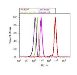
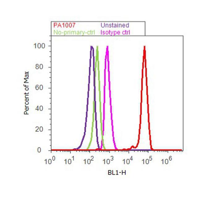
- Flow cytometry analysis of ER [p72] was done on Hep G2 cells. Cells were fixed with 70% ethanol for 10 minutes, permeabilized with 0.25% Triton™ X-100 for 20 minutes, and blocked with 5% BSA for 30 minutes at room temperature. Cells were labeled with ER [p72] Rabbit Polyclonal Antibody (PA1-007, red histogram) or with rabbit isotype control (pink histogram) at 3-5 ug/million cells in 2.5% BSA. After incubation at room temperature for 2 hours, the cells were labeled with Alexa Fluor® 488 Goat Anti-Rabbit Secondary Antibody (A11008) at a dilution of 1:400 for 30 minutes at room temperature. The representative 10, 000 cells were acquired and analyzed for each sample using an Attune® Acoustic Focusing Cytometer. The purple histogram represents unstained control cells and the green histogram represents no-primary-antibody control.
Supportive validation
- Submitted by
- Invitrogen Antibodies (provider)
- Main image
- Experimental details
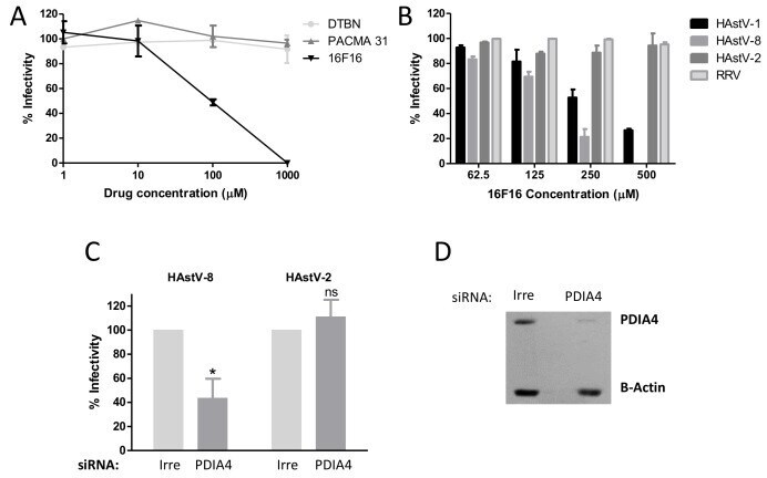
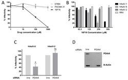
- Figure 4 PDIA4 is important for astrovirus infection. ( A ) Caco-2 cells were incubated with the indicated PDI inhibitors for 1 h at 37 degC, the cells were washed and infected with HAstV-8 and 16 h post-infection (hpi), and the viral infectivity was determined by an immunoperoxidase assay as described in Material and Methods. ( B ) Caco-2 cells were pre-treated for 1 h at 37 degC with the indicated concentrations of PDI inhibitor 16F16. Cells were washed and infected with either HAstV-1, -2, -8 or with rhesus rotavirus (RRV), and the viral infectivity was determined. Data in A and B are expressed as percentage of viral infectivity compared to control cells treated with DMSO, which was taken as 100%. ( C ) Caco-2 cells were transfected with an siRNA pool directed to PDIA4 or with an irrelevant (Irre) siRNA pool, and 72 h post-transfection (hpt) cells were infected with the indicated HAstV strain (MOI = 0.025). At 16 hpi, the cells were fixed, and the viral infectivity was determined as described previously. Virus infectivity is expressed as the percentage of infected cells obtained in the control-transfected cells (Irre), which was taken as 100%. The arithmetic mean +- SEM from three independent experiments performed in duplicate is shown. * p < 0.05. ( D ) A representative Western blot showing the effect of the siRNA-PDIA4 on the expression of PDIA4 as compared to cells transfected with an irrelevant siRNA. beta-Actin was used as a loading control.