 Explore
 Validate
 Learn
Learn Western blot
 Immunoprecipitation
Antibody data
- Antibody Data
- Antigen structure
- References [0]
- Comments [0]
- Validations
- Western blot [1]
- Immunohistochemistry [2]
- Other assay [1]
Submit
Validation data
Reference
Comment
Report error
- Product number
- PA5-118285 - Provider product page

- Provider
- Invitrogen Antibodies
- Product name
- HIP Polyclonal Antibody
- Antibody type
- Polyclonal
- Antigen
- Other
- Reactivity
- Human
- Host
- Rabbit
- Isotype
- IgG
- Vial size
- 100 µL
- Storage
- -20° C, Avoid Freeze/Thaw Cycles
No comments: Submit comment
Supportive validation
- Submitted by
- Invitrogen Antibodies (provider)
- Main image

- Experimental details
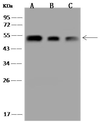
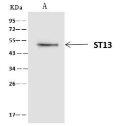
- Western Blot using HIP Polyclonal Antibody (Product # PA5-118285) at 1:500 dilution. Lane A: 293T Whole Cell Lysate, Lane B: SW480 Whole Cell Lysate, Lane C: RT4 Whole Cell Lysate. Lysates/proteins at 30 μg per lane. Secondary antibody: Goat Anti-Rabbit IgG (H+L)/HRP at 1:10,000 dilution. Developed using the ECL technique. Performed under reducing conditions. Predicted band size: 41 kDa. Observed band size: 50 kDa.
Supportive validation
- Submitted by
- Invitrogen Antibodies (provider)
- Main image
- Experimental details
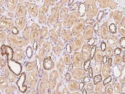
- Immunohistochemical staining of human HIP in human kidney with HIP Polyclonal Antibody (Product # PA5-118285, 1:300 dilution, formalin-fixed paraffin embedded sections).

- Submitted by
- Invitrogen Antibodies (provider)
- Main image
- Experimental details
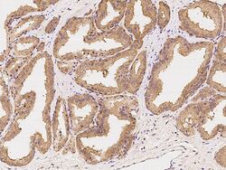
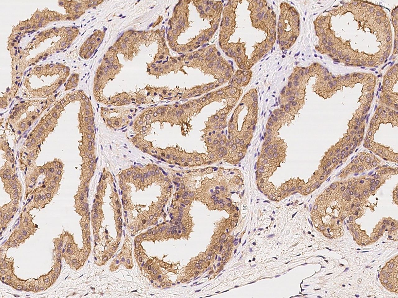
- Immunohistochemical staining of human HIP in human prostate with HIP Polyclonal Antibody (Product # PA5-118285, 1:300 dilution, formalin-fixed paraffin embedded sections).
Supportive validation
- Submitted by
- Invitrogen Antibodies (provider)
- Main image
- Experimental details
- HIP Immunoprecipitation using: Lane A: 0.5 mg 293T Whole Cell Lysate 4 µL with HIP Polyclonal Antibody (Product # PA5-118285) and 60 μg of Immunomagnetic beads Protein A/G. Primary antibody: HIP Polyclonal Antibody, at 1:100 dilution. Secondary antibody: Clean-Blot IP Detection Reagent (HRP) at 1:1,000dilution. Developed using the ECL technique. Performed under reducing conditions. Predicted band size: 41 kDa. Observed band size: 54 kDa.
