Explore
 Validate
 Learn
Learn Western blot
Antibody data
- Antibody Data
- Antigen structure
- References [0]
- Comments [0]
- Validations
- Western blot [3]
- Immunocytochemistry [5]
- Immunohistochemistry [3]
- Other assay [1]
Submit
Validation data
Reference
Comment
Report error
- Product number
- MA1-123 - Provider product page

- Provider
- Invitrogen Antibodies
- Product name
- RhoA/RhoC Monoclonal Antibody (1B3-4A10)
- Antibody type
- Monoclonal
- Antigen
- Recombinant full-length protein
- Description
- MA1-123 detects GTPases RhoA and RhoC, but does not react with RhoB.
- Antibody clone number
- 1B3-4A10
- Concentration
- 1 mg/mL
No comments: Submit comment
Supportive validation
- Submitted by
- Invitrogen Antibodies (provider)
- Main image
- Experimental details
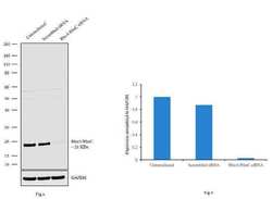
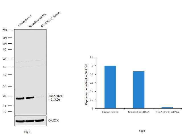
- Knockdown of RhoA/RhoC was achieved by transfecting SW-480 with RhoA and RhoC specific siRNAs (Silencer® select Product # s758, s97). Western blot analysis (Fig. a) was performed using membrane extracts from the RhoA and RhoC knockdown cells (lane 3), non-specific scrambled siRNA transfected cells (lane 2) and untransfected cells (lane 1). The blots were probed with RhoA/RhoC Monoclonal Antibody (1B3-4A10) (Product # MA1-123, 1:1000 dilution) and Goat anti-Mouse IgG (H+L) Superclonal™ Secondary Antibody, HRP conjugate (Product # A28177, 0.25 µg/mL, 1:4000 dilution). Densitometric analysis of this western blot is shown in histogram (Fig. b). Loss of signal upon siRNA mediated knock down confirms that antibody is specific to RhoA/RhoC.
- Submitted by
- Invitrogen Antibodies (provider)
- Main image
- Experimental details
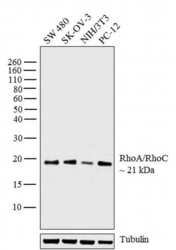
- Western blot analysis was performed on whole cell extracts (30 µg lysate) of SW 480 (Lane 1), SK-OV-3 (Lane 2), NIH/3T3 (Lane 3) and PC-12 (Lane 4). The blot was probed with Anti-RhoA/RhoC Monoclonal Antibody (Product # MA1-123, 1:1000 dilution) and detected by chemiluminescence using Goat anti-Mouse IgG (H+L) Superclonal™ Secondary Antibody, HRP conjugate (Product # A28177, 0.25 µg/mL, 1:4000 dilution). A 21 kDa band corresponding to RhoA/RhoC was observed across the cell lines tested.

- Submitted by
- Invitrogen Antibodies (provider)
- Main image
- Experimental details
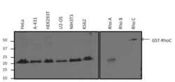
- Western blot analysis of RhoA/C was performed by loading 25 µg of various whole cell lysates or 0.5 µg of purified protein (RhoA, RhoB, and a GST-RhoC fusion) per well onto a 4-20% Tris-HCl polyacrylamide gel. Proteins were transferred to a PVDF membrane and blocked with 5% BSA/TBST for at least 1 hour. The membrane was probed with a RhoA/C monoclonal antibody (Product # MA1-123) at a dilution of 1:1000 overnight at 4°C on a rocking platform, washed in TBS-0.1%Tween-20, and probed with a goat anti-mouse IgG-HRP secondary antibody (Product # 31430) at a dilution of 1:15,000 for at least 1 hour. Chemiluminescent detection was performed using SuperSignal West Dura (Product # 34075).

Supportive validation
- Submitted by
- Invitrogen Antibodies (provider)
- Main image
- Experimental details
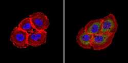
- Immunofluorescent analysis of Rho A/C (green) showing staining in the in the cytoplasm of A431 cells (right) compared to a negative control without primary antibody (left). Formalin-fixed cells were permeabilized with 0.1% Triton X-100 in TBS for 5-10 minutes and blocked with 3% BSA-PBS for 30 minutes at room temperature. Cells were probed with a Rho A/C monoclonal antibody (Product # MA1-123) in 3% BSA-PBS at a dilution of 1:100 and incubated overnight at 4ºC in a humidified chamber. Cells were washed with PBST and incubated with a DyLight-conjugated secondary antibody in PBS at room temperature in the dark. F-actin (red) was stained with a fluorescent red phalloidin and nuclei (blue) were stained with Hoechst or DAPI. Images were taken at a magnification of 60x.

- Submitted by
- Invitrogen Antibodies (provider)
- Main image
- Experimental details
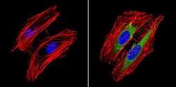
- Immunofluorescent analysis of Rho A/C (green) showing staining in the in the cytoplasm of Hela cells (right) compared to a negative control without primary antibody (left). Formalin-fixed cells were permeabilized with 0.1% Triton X-100 in TBS for 5-10 minutes and blocked with 3% BSA-PBS for 30 minutes at room temperature. Cells were probed with a Rho A/C monoclonal antibody (Product # MA1-123) in 3% BSA-PBS at a dilution of 1:100 and incubated overnight at 4ºC in a humidified chamber. Cells were washed with PBST and incubated with a DyLight-conjugated secondary antibody in PBS at room temperature in the dark. F-actin (red) was stained with a fluorescent red phalloidin and nuclei (blue) were stained with Hoechst or DAPI. Images were taken at a magnification of 60x.

- Submitted by
- Invitrogen Antibodies (provider)
- Main image
- Experimental details
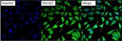
- Immunofluorescent analysis of RhoA/C (green) in HeLa cells. Formalin fixed cells were permeabilized with 0.1% Triton X-100 in TBS for 10 minutes at room temperature and blocked with 0.3%% BSA/TBST (Product # 37525) for 15 minutes at room temperature. Cells were probed with a RhoA/C monoclonal antibody (Product # MA1-123) at a dilution of 1:100 for at least 1 hour at room temperature, washed with PBS, and incubated with DyLight 488 goat anti-mouse IgG secondary antibody (Product # 35502) at a dilution of 1:400 for 30 minutes at room temperature. Nuclei (blue) were stained with Hoechst 33342 dye (Product # 62249). Images were taken on a Thermo Scientific ArrayScan at 20X magnification.

- Submitted by
- Invitrogen Antibodies (provider)
- Main image
- Experimental details
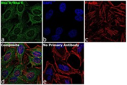
- Immunofluorescence analysis of RhoA/RhoC was performed using 70% confluent log phase HeLa cells. The cells were fixed with 4% paraformaldehyde for 10 minutes, permeabilized with 0.1% Triton™ X-100 for 15 minutes, and blocked with 1% BSA for 1 hour at room temperature. The cells were labeled with RhoA/RhoC Mouse Monoclonal Antibody (Product # MA1-123) at 5 µg/mL in 0.1% BSA, incubated at 4 degree Celsius overnight and then labeled with Goat anti-Mouse IgG (H+L) Superclonal™ Secondary Antibody, Alexa Fluor® 488 conjugate (Product # A28175) at a dilution of 1:2000 for 45 minutes at room temperature (Panel a: green). Nuclei (Panel b: blue) were stained with SlowFade® Gold Antifade Mountant with DAPI (Product # S36938). F-actin (Panel c: red) was stained with Rhodamine Phalloidin (Product # R415, 1:300). Panel d represents the merged image showing cytoplasmic localization. Panel e represents control cells with no primary antibody to assess background. The images were captured at 60X magnification.
- Submitted by
- Invitrogen Antibodies (provider)
- Main image
- Experimental details
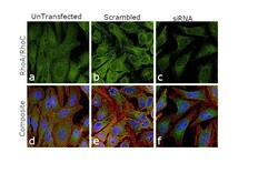
- Knockdown of RhoA/RhoC was achieved by transfecting HeLa cells with RhoA,RhoC specific siRNAs (Silencer® select Product # s758, s97). Immunofluorescence analysis was performed using untransfected HeLa cells (panels a, d), transfected with non-specific scrambled siRNA (panels b,e) and transfected with RhoA and RhoC specific siRNAs (panel c,f). Cells were fixed, permeabilized, and probed with RhoA/RhoC Monoclonal Antibody (1B3-4A10) (Product # MA1-123, 5 µg/mL), followed by labelling with Goat anti-Mouse IgG (H+L) Superclonal™ Secondary Antibody, Alexa Fluor 488 (Product # A28175, 1:2000). Nuclei (blue) were stained using ProLong™ Diamond Antifade Mountant with DAPI (Product # P36962) and Rhodamine Phalloidin (Product # R415, 1:300) was used for cytoskeletal F-actin (red) staining. Reduction of specific cytoplasmic localization was observed upon siRNA mediated knockdown (panel c,f) confirming specificity of the antibody to RhoA/RhoC. The images were captured at 60X magnification.
Supportive validation
- Submitted by
- Invitrogen Antibodies (provider)
- Main image
- Experimental details
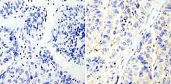
- Immunohistochemistry analysis of Rho A/C showing staining in the cytoplasm of paraffin-embedded human hepatocarcinoma (right) compared with a negative control without primary antibody (left). To expose target proteins, antigen retrieval was performed using 10mM sodium citrate (pH 6.0), microwaved for 8-15 min. Following antigen retrieval, tissues were blocked in 3% H2O2-methanol for 15 min at room temperature, washed with ddH2O and PBS, and then probed with a Rho A/C monoclonal antibody (Product # MA1-123) diluted in 3% BSA-PBS at a dilution of 1:200 overnight at 4°C in a humidified chamber. Tissues were washed extensively in PBST and detection was performed using an HRP-conjugated secondary antibody followed by colorimetric detection using a DAB kit. Tissues were counterstained with hematoxylin and dehydrated with ethanol and xylene to prep for mounting.

- Submitted by
- Invitrogen Antibodies (provider)
- Main image
- Experimental details
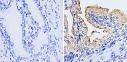
- Immunohistochemistry analysis of Rho A/C showing staining in the cytoplasm and membrane of paraffin-embedded human prostate carcinoma (right) compared with a negative control without primary antibody (left). To expose target proteins, antigen retrieval was performed using 10mM sodium citrate (pH 6.0), microwaved for 8-15 min. Following antigen retrieval, tissues were blocked in 3% H2O2-methanol for 15 min at room temperature, washed with ddH2O and PBS, and then probed with a Rho A/C monoclonal antibody (Product # MA1-123) diluted in 3% BSA-PBS at a dilution of 1:200 overnight at 4°C in a humidified chamber. Tissues were washed extensively in PBST and detection was performed using an HRP-conjugated secondary antibody followed by colorimetric detection using a DAB kit. Tissues were counterstained with hematoxylin and dehydrated with ethanol and xylene to prep for mounting.

- Submitted by
- Invitrogen Antibodies (provider)
- Main image

- Experimental details
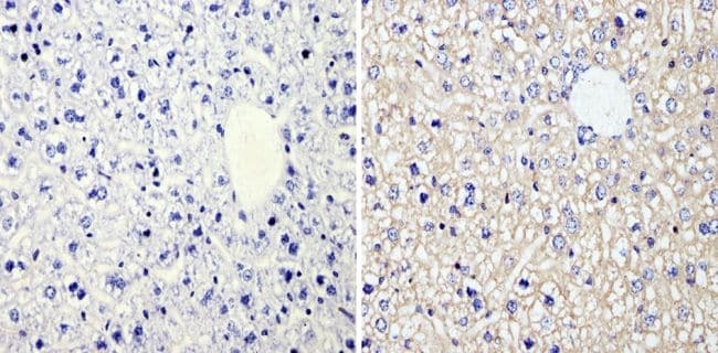
- Immunohistochemistry analysis of Rho A/C showing staining in the cytoplasm and membrane of paraffin-embedded mouse liver tissue (right) compared with a negative control without primary antibody (left). To expose target proteins, antigen retrieval was performed using 10mM sodium citrate (pH 6.0), microwaved for 8-15 min. Following antigen retrieval, tissues were blocked in 3% H2O2-methanol for 15 min at room temperature, washed with ddH2O and PBS, and then probed with a Rho A/C monoclonal antibody (Product # MA1-123) diluted in 3% BSA-PBS at a dilution of 1:100 overnight at 4°C in a humidified chamber. Tissues were washed extensively in PBST and detection was performed using an HRP-conjugated secondary antibody followed by colorimetric detection using a DAB kit. Tissues were counterstained with hematoxylin and dehydrated with ethanol and xylene to prep for mounting.
Supportive validation
- Submitted by
- Invitrogen Antibodies (provider)
- Main image

- Experimental details
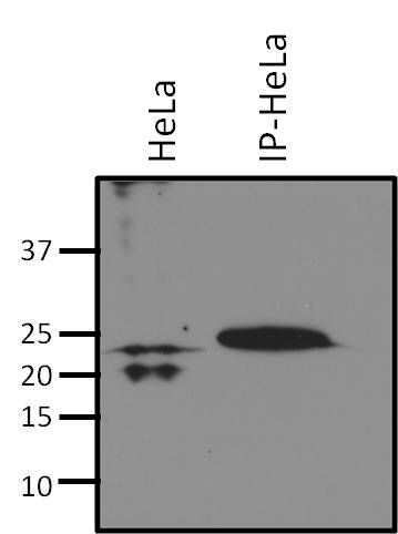
- Immunoprecipitation of RhoA/C was performed on HeLa cells. Antigen-antibody complexes were formed by incubating 500 µg of whole cell lysate with 2 µg of a RhoA/C monoclonal antibody (Product # MA1-123) overnight on a rocking platform at 4øC. The immune complexes were captured on 50 µL Protein A/G Plus Agarose (Product # 20423), washed extensively, and eluted with Lane Marker Reducing Sample Buffer (Product # 39000). HeLa cell lysate was run as a control (left lane). Samples were resolved on a 4-20% Tris-HCl polyacrylamide gel, transferred to PVDF membrane, and blocked with 5% BSA/TBS-0.1%Tween for at least 1 hour. The membrane was probed with a RhoA/C monoclonal antibody (Product # MA1-123) at a dilution of 1:1000 overnight at 4øC, washed in TBST, and probed with Clean-Blot IP Detection Reagent (Product # 21230) at a dilution of 1:2500 for at least 1 hour. Chemiluminescent detection was performed using SuperSignal West Dura (Product # 34075).