 Explore
 Validate
 Learn
LearnHPA001667
antibody from Atlas Antibodies
Targeting: PAPPA
ASBABP2, DIPLA1, IGFBP-4ase, PAPA, PAPP-A, PAPPA1
 Immunohistochemistry
Antibody data
- Antibody Data
- Antigen structure
- References [0]
- Comments [0]
- Validations
- Immunohistochemistry [6]
Submit
Validation data
Reference
Comment
Report error
- Product number
- HPA001667 - Provider product page

- Provider
- Atlas Antibodies
- Proper citation
- Atlas Antibodies Cat#HPA001667, RRID:AB_1854960
- Product name
- Anti-PAPPA
- Antibody type
- Polyclonal
- Reactivity
- Human
- Host
- Rabbit
- Conjugate
- Unconjugated
- Antigen sequence
SCLDHNSESIILPMNVTVRDIPHWLNPTRVERVVC
TAGLKWYPHPALIHCVKGCEPFMGDNYCDAINNRA
FCNYDGGDCCTSTVKTKKVTPFPMSCDLQGDCACR
DPQAQEHSRKDL- Isotype
- IgG
- Vial size
- 100 µl
- Storage
- Store at +4°C for short term storage. Long time storage is recommended at -20°C.
No comments: Submit comment
Enhanced validation
Supportive validation
- Submitted by
- Atlas Antibodies (provider)
- Enhanced method
- Orthogonal validation
- Main image

- Experimental details
- Immunohistochemistry analysis in human placenta and skeletal muscle tissues using HPA001667 antibody. Corresponding PAPPA RNA-seq data are presented for the same tissues.
- Sample type
- HUMAN

Supportive validation
- Submitted by
- Atlas Antibodies (provider)
- Main image

- Experimental details
- Immunohistochemical staining of human placenta shows strong cytoplasmic positivity in trophoblastic cells.

- Submitted by
- Atlas Antibodies (provider)
- Main image

- Experimental details
- Immunohistochemical staining of human placenta shows moderate to strong cytoplasmic positivity in trophoblastic cells.
- Sample type
- HUMAN

- Submitted by
- Atlas Antibodies (provider)
- Main image
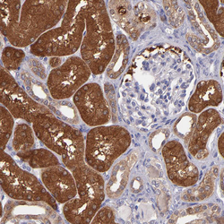
- Experimental details
- Immunohistochemical staining of human kidney shows moderate to strong cytoplasmic positivity in cells in tubules.
- Sample type
- HUMAN

- Submitted by
- Atlas Antibodies (provider)
- Main image

- Experimental details
- Immunohistochemical staining of human endometrium shows moderate to strong cytoplasmic positivity in glandular cells.
- Sample type
- HUMAN

- Submitted by
- Atlas Antibodies (provider)
- Main image

- Experimental details
- Immunohistochemical staining of human skeletal muscle shows no cytoplasmic positivity in myocytes as expected.
- Sample type
- HUMAN
