 Explore
 Validate
 Learn
Learn Western blot
Antibody data
- Antibody Data
- Antigen structure
- References [4]
- Comments [0]
- Validations
- Western blot [3]
- Immunocytochemistry [2]
- Immunohistochemistry [2]
- Other assay [3]
Submit
Validation data
Reference
Comment
Report error
- Product number
- PA5-34787 - Provider product page

- Provider
- Invitrogen Antibodies
- Product name
- Collagen III Polyclonal Antibody
- Antibody type
- Polyclonal
- Antigen
- Recombinant protein fragment
- Description
- Recommended positive controls: HeLa, SK-N-SH, SK-N-AS, mouse brain, rat brain. Predicted reactivity: Mouse (92%), Rat (92%), Pig (93%), Chicken (86%), Rhesus Monkey (97%), Bovine (93%). IHC notes, Requires antigen retrieval using heat mediated 10mM Citrate buffer (pH6.0) or Tris-EDTA buffer (pH8.0) Store product as a concentrated solution. Centrifuge briefly prior to opening the vial.
- Reactivity
- Human
- Host
- Rabbit
- Isotype
- IgG
- Vial size
- 100 µL
- Concentration
- 1.98 mg/mL
- Storage
- Store at 4°C short term. For long term storage, store at -20°C, avoiding freeze/thaw cycles.
Submitted references Dystrophin Deficiency Causes Progressive Depletion of Cardiovascular Progenitor Cells in the Heart.
Translation of Tudor-SN, a novel terminal oligo-pyrimidine (TOP) mRNA, is regulated by the mTORC1 pathway in cardiomyocytes.
3D Bioprinting of Human Adipose-Derived Stem Cells and Their Tenogenic Differentiation in Clinical-Grade Medium.
Exogenous supply of Hsp47 triggers fibrillar collagen deposition in skin cell cultures in vitro.
Jelinkova S, Sleiman Y, Fojtík P, Aimond F, Finan A, Hugon G, Scheuermann V, Beckerová D, Cazorla O, Vincenti M, Amedro P, Richard S, Jaros J, Dvorak P, Lacampagne A, Carnac G, Rotrekl V, Meli AC
International journal of molecular sciences 2021 May 10;22(9)
International journal of molecular sciences 2021 May 10;22(9)
Translation of Tudor-SN, a novel terminal oligo-pyrimidine (TOP) mRNA, is regulated by the mTORC1 pathway in cardiomyocytes.
Gan S, Su C, Ma J, Liu M, Cui X, Xin L, Ren Y, Gao X, Ge L, Wei M, Yang J
RNA biology 2021 Jun;18(6):900-913
RNA biology 2021 Jun;18(6):900-913
3D Bioprinting of Human Adipose-Derived Stem Cells and Their Tenogenic Differentiation in Clinical-Grade Medium.
Stanco D, Boffito M, Bogni A, Puricelli L, Barrero J, Soldati G, Ciardelli G
International journal of molecular sciences 2020 Nov 18;21(22)
International journal of molecular sciences 2020 Nov 18;21(22)
Exogenous supply of Hsp47 triggers fibrillar collagen deposition in skin cell cultures in vitro.
Khan ES, Sankaran S, Llontop L, Del Campo A
BMC molecular and cell biology 2020 Mar 30;21(1):22
BMC molecular and cell biology 2020 Mar 30;21(1):22
No comments: Submit comment
Supportive validation
- Submitted by
- Invitrogen Antibodies (provider)
- Main image
- Experimental details
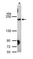
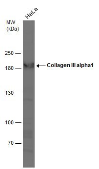
- Western blot analysis of COL3A1/Collagen III using 30 µg of HeLa lysate. Samples were loaded onto a 5% SDS-PAGE gel and probed with a COL3A1/Collagen III polyclonal antibody (Product # PA5-34787) at a dilution of 1:1000.

- Submitted by
- Invitrogen Antibodies (provider)
- Main image
- Experimental details
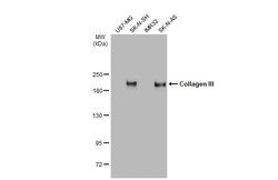
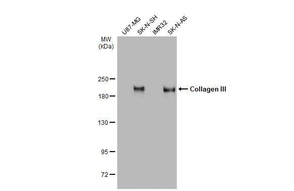
- Western Blot analysis of Collagen III was performed by separating 30 µg of various whole cell extracts by 5% SDS-PAGE. Proteins were transferred to a membrane and probed with a Collagen III Polyclonal Antibody (Product # PA5-34787) at a dilution of 1:2000 and a HRP-conjugated anti-rabbit IgG secondary antibody.
- Submitted by
- Invitrogen Antibodies (provider)
- Main image
- Experimental details
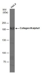
- Western Blot analysis of Collagen III was performed by separating 30 µg of whole cell extract by 5% SDS-PAGE. Proteins were transferred to a membrane and probed with a Collagen III Polyclonal Antibody (Product # PA5-34787) at a dilution of 1:1000.
Supportive validation
- Submitted by
- Invitrogen Antibodies (provider)
- Main image

- Experimental details
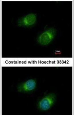
- Immunofluorescent analysis of COL3A1/Collagen III in methanol-fixed HeLa cells using a COL3A1/Collagen III polyclonal antibody (Product # PA5-34787) at a 1:50 dilution.
- Submitted by
- Invitrogen Antibodies (provider)
- Main image
- Experimental details
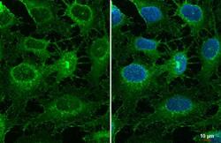
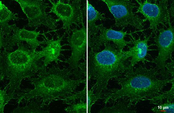
- Collagen III Polyclonal Antibody detects Collagen III protein at cell membrane by immunofluorescent analysis. Sample: HeLa cells were fixed in ice-cold MeOH for 5 min. Green: Collagen III stained by Collagen III Polyclonal Antibody (Product # PA5-34787) diluted at 1:500.
Supportive validation
- Submitted by
- Invitrogen Antibodies (provider)
- Main image

- Experimental details
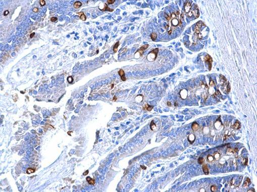
- Collagen III Polyclonal Antibody detects Collagen III protein at secreted on mouse intestine by immunohistochemical analysis. Sample: Paraffin-embedded mouse intestine. Collagen III Polyclonal Antibody (Product # PA5-34787) dilution: 1:500. Antigen Retrieval: EDTA based buffer, pH 8.0, 15 min.
- Submitted by
- Invitrogen Antibodies (provider)
- Main image
- Experimental details
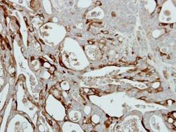
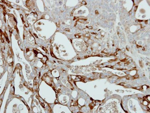
- Immunohistochemical analysis of paraffin-embedded NCIN87 xenograft, using Collagen III (Product # PA5-34787) antibody at 1:500 dilution. Antigen Retrieval: EDTA based buffer, pH 8.0, 15 min.
Supportive validation
- Submitted by
- Invitrogen Antibodies (provider)
- Main image
- Experimental details
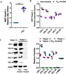
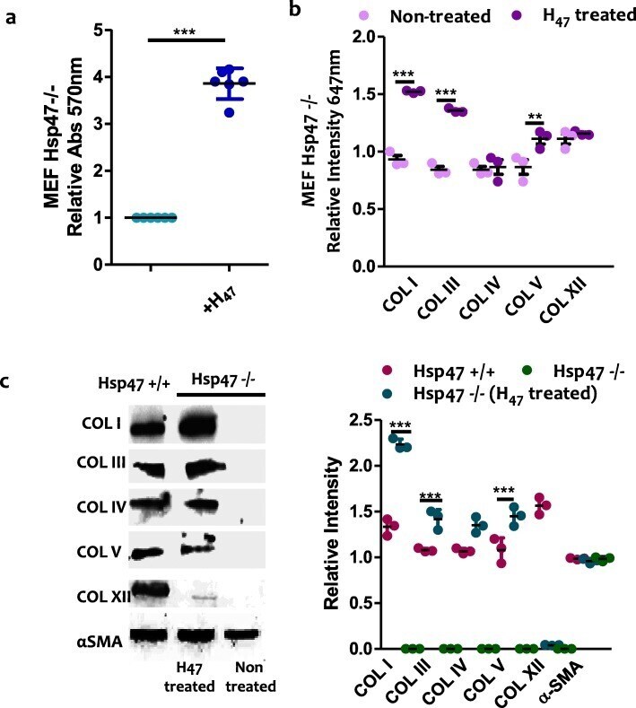
- Fig. 5 a. Quantification of fibrillar collagen deposited in MEF Hsp47-/- and Hsp47 +/+cells (control) at 24 h after H 47 treatment (0.5 muM) using Sirius Red assay. b. Quantification of deposited COL I, III, IV, V and XII from immuno staining assays in MEF Hsp47-/- cultures. Error bars representing standard deviation from n-3 experiments in a and c , The plots in both a and c assays were normalized by MEF Hsp47-/- cells untreated condition taken as 1. Statistical significance in a and c was analyzed by Tukey test. Significance was calculated by comparing non treated against Hsp47 treated cells (mean +- SD, ANOVA, *** p < 0.001). c. Western blot of COL I, III, IV, V and XII in deposited collagen from MEF Hsp47 +/+, Hsp47 -/- and Hsp47-/- cultures 24 h after treatment with 0.5 muM H 47 . Equal amount of cells were analyzed. Black bands indicate signal from collagen subtypes specific antibody. The wiskers plot indicates relative gel bands intensity of different collagen types in MEF Hsp47 +/+, Hsp47 -/- and Hsp47-/- H 47 treated conditions. Error bar indicates standard deviation from n-3 experiments. Statistical significance was analyzed by Tukey test. Significance was calculated by comparing non treated Hsp47 -/- against Hsp47 treated cells (mean +- SD, ANOVA, *** p < 0.001)
- Submitted by
- Invitrogen Antibodies (provider)
- Main image
- Experimental details
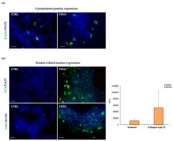
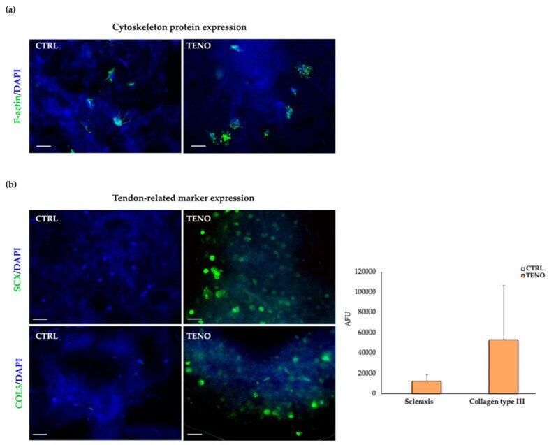
- Figure 7 Cytoskeleton- and tendon-related marker expression: ( a ) representative images of both CTRL- and TENO-cultured ASCs embedded in NFC/A scaffolds stained at day 14 of culture with phalloidin (green) for F-actin detection and counterstained with DAPI to visualize cell nuclei (blue) (32x magnifications; 10um scale bar); ( b ) on the left, representative images of both CTRL- and TENO-cultured ASC constructs stained with scleraxis (SCX) and collagen type III (COL3) at days 3 and 14 of differentiation, respectively, visualized with Alexa Fluor 488 (green) and counterstained with DAPI (blue) (20x magnification; 10um scale bar); on the right, the level of cellular fluorescence intensity from fluorescence microscopy images ( N = 3) expressed as average +- sem of arbitrary fluorescence units (AFU).
- Submitted by
- Invitrogen Antibodies (provider)
- Main image
- Experimental details
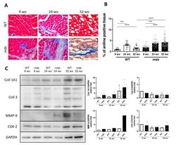
- Figure 4 Fibrotic deposit and cardiac dysfunction correlate with decreasing CVPC presence in mdx heart. ( A ) Representative images of histological analysis stained using Masson trichrome technique showing myocytes (in red) and collagenous fibrotic tissue (in blue) in the left ventricle of WT and mdx hearts at 9, 24, and 52 wo. Line represents 100 um. ( B ) The ratio of red and blue stained tissue was evaluated in WT hearts (open bars and black dots, n = 4-11 slices/3 animals per group) and mdx hearts (black bars and grey dots, n = 3-16 slices/3 animals per group) at the age of 24 wo and further at 52 wo. Statistical significance was calculated by Kruskal-Wallis test and Dunn's multiple comparison post-hoc test (*** p < 0.001, **** p < 0.0001). ( C ) Western blot analysis of collagen proteins and inflammatory proteins in the cardiac tissues. Left panel shows representative images of collagen 1A1 (Coll 1A1), collagen 3 (Coll 3), cyclooxygenase 2 (COX-2), and matrix metalloproteinase 9 (MMP-9) compared to the GAPDH control. The right panels show the normalized densitometry of each protein normalized by GAPDH content of WT (open bars, n = 2 animals) and mdx (black bars, n = 2 animals).
