Explore
 Validate
 Learn
Learn Western blot
Antibody data
- Antibody Data
- Antigen structure
- References [0]
- Comments [0]
- Validations
- Western blot [2]
- Immunocytochemistry [1]
- Immunohistochemistry [1]
Submit
Validation data
Reference
Comment
Report error
- Product number
- NBP2-30036 - Provider product page

- Provider
- Novus Biologicals
- Product name
- Rabbit Polyclonal PCYT1A Antibody
- Antibody type
- Polyclonal
- Description
- Protein A purified.
- Reactivity
- Human
- Host
- Rabbit
- Isotype
- IgG
- Vial size
- 0.4 ml
- Concentration
- 0.5 mg/ml
- Storage
- Store at 4C short term. Aliquot and store at -20C long term. Avoid freeze-thaw cycles.
No comments: Submit comment
Supportive validation
- Submitted by
- Novus Biologicals (provider)
- Main image

- Experimental details
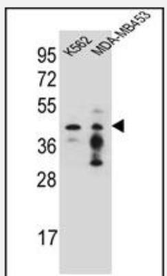
- Western Blot: PCYT1A Antibody [NBP2-30036] - Western blot analysis in K562,MDA-MB453 cell line lysates (35ug/lane).This demonstrates the PCYT1A antibody detected the PCYT1A protein (arrow).
- Submitted by
- Novus Biologicals (provider)
- Main image
- Experimental details
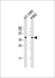
- Western Blot: PCYT1A Antibody [NBP2-30036] - All lanes : Anti- (N-term) at 1:1000 dilution Lane 1: HT-1080 whole cell lysate Lane 2: K562 whole cell lysate Lysates/proteins at 20 ug per lane. Secondary Goat Anti-Rabbit IgG, (H+L), Peroxidase conjugated at 1/10000 dilution. Predicted band size : 42 kDa Blocking/Dilution buffer: 5% NFDM/TBST.

Supportive validation
- Submitted by
- Novus Biologicals (provider)
- Main image

- Experimental details
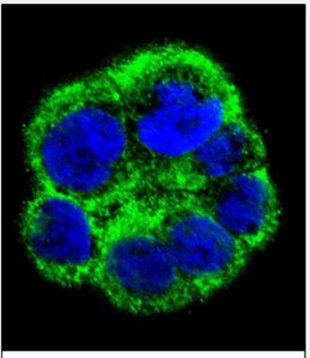
- Immunocytochemistry/Immunofluorescence: PCYT1A Antibody [NBP2-30036] - Confocal immunofluorescent analysis of (N-term)(NBP2-30036) with WiDr cell followed by Alexa Fluor 488-conjugated goat anti-rabbit lgG (green). DAPI was used to stain the cell nuclear (blue).
Supportive validation
- Submitted by
- Novus Biologicals (provider)
- Main image
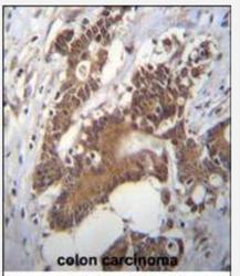
- Experimental details
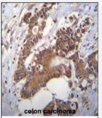
- Immunohistochemistry-Paraffin: PCYT1A Antibody [NBP2-30036] - Immunohistochemistry analysis in formalin fixed and paraffin embedded human colon carcinoma followed by peroxidase conjugation of the secondary antibody and DAB staining.This data demonstrates the use of (N-term) for immunohistochemistry. Clinical relevance has not been evaluated.