Explore
 Validate
 Learn
Learn Western blot
Antibody data
- Antibody Data
- Antigen structure
- References [1]
- Comments [0]
- Validations
- Western blot [1]
- Immunocytochemistry [1]
Submit
Validation data
Reference
Comment
Report error
- Product number
- AF7217 - Provider product page

- Provider
- R&D Systems
- Product name
- Human/Mouse RNF168 Antibody
- Antibody type
- Polyclonal
- Description
- Antigen Affinity-purified. Detects recombinant mouse RNF168 and recombinant human RNF168 in direct ELISAs and Western blots.
- Reactivity
- Human, Mouse
- Host
- Sheep
- Conjugate
- Unconjugated
- Antigen sequence
Q80XJ2- Isotype
- IgG
- Vial size
- 100 ug
- Concentration
- LYOPH
- Storage
- Use a manual defrost freezer and avoid repeated freeze-thaw cycles. 12 months from date of receipt, -20 to -70 °C as supplied. 1 month, 2 to 8 °C under sterile conditions after reconstitution. 6 months, -20 to -70 °C under sterile conditions after reconstitution.
Submitted references RNF168 and USP10 regulate topoisomerase IIα function via opposing effects on its ubiquitylation.
Guturi KKN, Bohgaki M, Bohgaki T, Srikumar T, Ng D, Kumareswaran R, El Ghamrasni S, Jeon J, Patel P, Eldin MS, Bristow R, Cheung P, Stewart GS, Raught B, Hakem A, Hakem R
Nature communications 2016 Aug 25;7:12638
Nature communications 2016 Aug 25;7:12638
No comments: Submit comment
Supportive validation
- Submitted by
- R&D Systems (provider)
- Main image
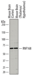
- Experimental details
- Detection of Mouse RNF168 by Western Blot. Western blot shows lysates of mouse brain (cortex) tissue and mouse brain (thalamus/hypothalamus) tissue. PVDF membrane was probed with 0.5 µg/mL of Sheep Anti-Mouse RNF168 Antigen Affinity-purified Polyclonal Antibody (Catalog # AF7217) followed by HRP-conjugated Anti-Sheep IgG Secondary Antibody (Catalog # HAF016). A specific band was detected for RNF168 at approximately 65 kDa (as indicated). This experiment was conducted under reducing conditions and using Immunoblot Buffer Group 1.

Supportive validation
- Submitted by
- R&D Systems (provider)
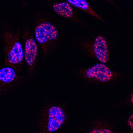
- Main image

- Experimental details
- RNF168 in HeLa Human Cell Line. RNF168 was detected in immersion fixed HeLa human cervical epithelial carcinoma cell line using Sheep Anti-Mouse RNF168 Antigen Affinity-purified Polyclonal Antibody (Catalog # AF7217) at 10 µg/mL for 3 hours at room temperature. Cells were stained using the NorthernLights™ 557-conjugated Anti-Sheep IgG Secondary Antibody (red; Catalog # NL010) and counterstained with DAPI (blue). View our protocol for Fluorescent ICC Staining of Cells on Coverslips.