Antibody data
- Antibody Data
- Antigen structure
- References [2]
- Comments [0]
- Validations
- Western blot [1]
- Immunocytochemistry [1]
- Immunohistochemistry [9]
- Other assay [1]
Submit
Validation data
Reference
Comment
Report error
- Product number
- PA5-104554 - Provider product page

- Provider
- Invitrogen Antibodies
- Product name
- Phospho-JAK1 (Tyr1034) Polyclonal Antibody
- Antibody type
- Polyclonal
- Antigen
- Synthetic peptide
- Description
- Antibody detects endogenous levels of JAK1 only when phosphorylated at Tyr1034, which site historically referenced as Tyr1022.
- Reactivity
- Human, Mouse, Rat
- Host
- Rabbit
- Isotype
- IgG
- Vial size
- 100 µL
- Concentration
- 1 mg/mL
- Storage
- -20°C
Submitted references Tofacitinib Ameliorates Retinal Vascular Leakage in a Murine Model of Diabetic Retinopathy with Type 2 Diabetes.
IL-17A Damages the Blood-Retinal Barrier through Activating the Janus Kinase 1 Pathway.
Byrne EM, Llorián-Salvador M, Lyons TJ, Chen M, Xu H
International journal of molecular sciences 2021 Nov 2;22(21)
International journal of molecular sciences 2021 Nov 2;22(21)
IL-17A Damages the Blood-Retinal Barrier through Activating the Janus Kinase 1 Pathway.
Byrne EM, Llorián-Salvador M, Tang M, Margariti A, Chen M, Xu H
Biomedicines 2021 Jul 16;9(7)
Biomedicines 2021 Jul 16;9(7)
No comments: Submit comment
Supportive validation
- Submitted by
- Invitrogen Antibodies (provider)
- Main image

- Experimental details
- Western blot analysis of Phospho-JAK1 (Tyr1034, Tyr1035) in MCF7 cells treated with IFN-alpha1 (left lane: treated with blocking peptide). Samples were incubated with Phospho-JAK1 (Tyr1034, Tyr1035) polyclonal antibody (Product # PA5-104554).
Supportive validation
- Submitted by
- Invitrogen Antibodies (provider)
- Main image
- Experimental details
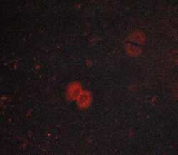
- Immunofluorescent analysis of Phospho-JAK1 (Tyr1034, Tyr1035) in lovo cells. Samples were fixed with paraformaldehyde, permeabilized with 0.1% saponin, blocked with 10% serum (45 min, 37°C) incubated with Phospho-JAK1 (Tyr1034, Tyr1035) polyclonal antibody (Product # PA5-104554) using a dilution of 1:400 (1 hr, 37°C), and followed by goat anti-rabbit IgG Alexa Fluor 594 at a dilution of 1:600.
Supportive validation
- Submitted by
- Invitrogen Antibodies (provider)
- Main image

- Experimental details
- Immunohistochemistry analysis of paraffin-embedded Phospho-JAK1 (Tyr1034, Tyr1035) in human appendiceal tissue sections. Antigen retrieval was performed using citrate buffer. Samples were blocked with blocking buffer (1.5 hr, 22°C), incubated with Phospho-JAK1 (Tyr1034, Tyr1035) polyclonal antibody (Product # PA5-104554) using a dilution of 1:100 (1.5 hr, 22°C), followed by HRP conjugated goat anti-rabbit.
- Submitted by
- Invitrogen Antibodies (provider)
- Main image
- Experimental details
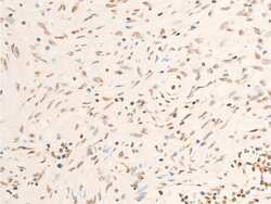
- Immunohistochemistry analysis of paraffin-embedded Phospho-JAK1 (Tyr1034, Tyr1035) in human muscle tumor tissue sections. Antigen retrieval was performed using citrate buffer. Samples were blocked with blocking buffer (1.5 hr, 22°C), incubated with Phospho-JAK1 (Tyr1034, Tyr1035) polyclonal antibody (Product # PA5-104554) using a dilution of 1:100 (1.5 hr, 22°C), followed by HRP conjugated goat anti-rabbit.
- Submitted by
- Invitrogen Antibodies (provider)
- Main image

- Experimental details
- Immunohistochemistry analysis of paraffin-embedded Phospho-JAK1 (Tyr1034, Tyr1035) in human seminoma tissue sections. Antigen retrieval was performed using citrate buffer. Samples were blocked with blocking buffer (1.5 hr, 22°C), incubated with Phospho-JAK1 (Tyr1034, Tyr1035) polyclonal antibody (Product # PA5-104554) using a dilution of 1:100 (1.5 hr, 22°C), followed by HRP conjugated goat anti-rabbit.
- Submitted by
- Invitrogen Antibodies (provider)
- Main image

- Experimental details
- Immunohistochemistry analysis of paraffin-embedded Phospho-JAK1 (Tyr1034, Tyr1035) in mouse intestinal tissue sections. Antigen retrieval was performed using citrate buffer. Samples were blocked with blocking buffer (1.5 hr, 22°C), incubated with Phospho-JAK1 (Tyr1034, Tyr1035) polyclonal antibody (Product # PA5-104554) using a dilution of 1:100 (1.5 hr, 22°C), followed by HRP conjugated goat anti-rabbit.
- Submitted by
- Invitrogen Antibodies (provider)
- Main image

- Experimental details
- Immunohistochemistry analysis of paraffin-embedded Phospho-JAK1 (Tyr1034, Tyr1035) in mouse lung tissue sections. Antigen retrieval was performed using citrate buffer. Samples were blocked with blocking buffer (1.5 hr, 22°C), incubated with Phospho-JAK1 (Tyr1034, Tyr1035) polyclonal antibody (Product # PA5-104554) using a dilution of 1:100 (1.5 hr, 22°C), followed by HRP conjugated goat anti-rabbit.
- Submitted by
- Invitrogen Antibodies (provider)
- Main image

- Experimental details
- Immunohistochemistry analysis of paraffin-embedded Phospho-JAK1 (Tyr1034, Tyr1035) in mouse testis tissue sections. Antigen retrieval was performed using citrate buffer. Samples were blocked with blocking buffer (1.5 hr, 22°C), incubated with Phospho-JAK1 (Tyr1034, Tyr1035) polyclonal antibody (Product # PA5-104554) using a dilution of 1:100 (1.5 hr, 22°C), followed by HRP conjugated goat anti-rabbit.
- Submitted by
- Invitrogen Antibodies (provider)
- Main image
- Experimental details
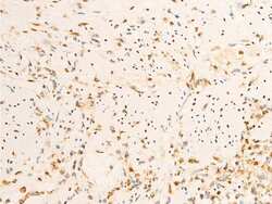
- Immunohistochemistry analysis of paraffin-embedded Phospho-JAK1 (Tyr1034, Tyr1035) in rat appendiceal tissue sections. Antigen retrieval was performed using citrate buffer. Samples were blocked with blocking buffer (1.5 hr, 22°C), incubated with Phospho-JAK1 (Tyr1034, Tyr1035) polyclonal antibody (Product # PA5-104554) using a dilution of 1:100 (1.5 hr, 22°C), followed by HRP conjugated goat anti-rabbit.
- Submitted by
- Invitrogen Antibodies (provider)
- Main image
- Experimental details
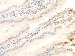
- Immunohistochemistry analysis of paraffin-embedded Phospho-JAK1 (Tyr1034, Tyr1035) in rat intestinal tissue sections. Antigen retrieval was performed using citrate buffer. Samples were blocked with blocking buffer (1.5 hr, 22°C), incubated with Phospho-JAK1 (Tyr1034, Tyr1035) polyclonal antibody (Product # PA5-104554) using a dilution of 1:100 (1.5 hr, 22°C), followed by HRP conjugated goat anti-rabbit.
- Submitted by
- Invitrogen Antibodies (provider)
- Main image

- Experimental details
- Immunohistochemistry analysis of paraffin-embedded Phospho-JAK1 (Tyr1034, Tyr1035) in rat uterine tissue sections. Antigen retrieval was performed using citrate buffer. Samples were blocked with blocking buffer (1.5 hr, 22°C), incubated with Phospho-JAK1 (Tyr1034, Tyr1035) polyclonal antibody (Product # PA5-104554) using a dilution of 1:100 (1.5 hr, 22°C), followed by HRP conjugated goat anti-rabbit.
Supportive validation
- Submitted by
- Invitrogen Antibodies (provider)
- Main image
- Experimental details
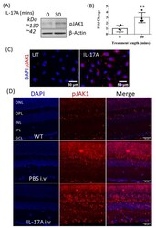
- Figure 3 The effect of IL-17A on pJAK1 expression in bEnd.3 cells and the WT mouse retina. JAK1 phosphorylation was examined in bEnd.3 cells by Western blot and immunostaining, and in mouse retinal sections 48 h after IL-17A i.v. administration. ( A ) Representative Western blot and corresponding densitometry of pJAK1 in control (0 min) and IL-17A-treated bEnd3 cells. ( B ) Quantification of pJAK1 expression detected by Western blotting. Mean +- SD; ** p = 4. ( C ) Representative images of pJAK1 immunocytochemistry in bEnd.3 cells from n >= 3 independent experiments. ( D ) Immunostaining of pJAK1 expression (red) in the murine retina 48 h after IL-17A or PBS i.v. injection compared to control non-injected wild-type (WT) mice, n >= 4.
 Explore
 Validate
 Learn
Learn Western blot