 Explore
 Validate
 Learn
Learn Western blot
Antibody data
- Antibody Data
- Antigen structure
- References [23]
- Comments [0]
- Validations
- Western blot [1]
- Immunocytochemistry [1]
- Flow cytometry [1]
- Other assay [8]
Submit
Validation data
Reference
Comment
Report error
- Product number
- 44-804G - Provider product page

- Provider
- Invitrogen Antibodies
- Product name
- Phospho-IR/IGF1R (Tyr1162, Tyr1163) Polyclonal Antibody
- Antibody type
- Polyclonal
- Antigen
- Synthetic peptide
- Description
- The antibody has been negatively preadsorbed using a non-phosphopeptide corresponding to the site of phosphorylation to remove antibody reactive with non-phosphorylated insulin/insulin-like growth factor-1 receptor (IR/IGF1R). The final product is generated by affinity chromatography using an IR/IGF1R-derived peptide that is phosphorylated at tyrosines 1162 and 1163 (tyrosines 1135 and 1136 for IGF1R. The antiserum was produced against a chemically synthesized phosphopeptide derived from the region of IR/IGF1R that contains tyrosines 1162 and 1163 of the human insulin receptor (IR) as numbered according to Ebina, et al. (1150 and 1151 according to Ullrich et al.). The corresponding residues in the IGF1R are 1135 and 1136. The sequence is conserved in mouse and rat for both the IR and IGF1R. Although exhibiting a preference for IR/IGF1R, this antibody has been shown by both peptide competition and protein blotting to react with other dual-phosphotyrosine motifs from proteins such as c-Met and Shc.
Submitted references The inhibitory effect of ECG and EGCG dimeric procyanidins on colorectal cancer cells growth is associated with their actions at lipid rafts and the inhibition of the epidermal growth factor receptor signaling.
Endocrine-Exocrine Signaling Drives Obesity-Associated Pancreatic Ductal Adenocarcinoma.
IGF2 Autocrine-Mediated IGF1R Activation Is a Clinically Relevant Mechanism of Osimertinib Resistance in Lung Cancer.
Lipid and glucose metabolism in hepatocyte cell lines and primary mouse hepatocytes: a comprehensive resource for in vitro studies of hepatic metabolism.
Ablation of Grb10 Specifically in Muscle Impacts Muscle Size and Glucose Metabolism in Mice.
Diabetes reversal by inhibition of the low-molecular-weight tyrosine phosphatase.
Epigenetic regulation of macrophage polarization and inflammation by DNA methylation in obesity.
Agonistic aptamer to the insulin receptor leads to biased signaling and functional selectivity through allosteric modulation.
The insulin and IGF1 receptor kinase domains are functional dimers in the activated state.
Targeting the insulin-like growth factor receptor and Src signaling network for the treatment of non-small cell lung cancer.
Hepatocyte Nicotinamide Adenine Dinucleotide Phosphate Reduced Oxidase 4 Regulates Stress Signaling, Fibrosis, and Insulin Sensitivity During Development of Steatohepatitis in Mice.
Methionine restriction restores a younger metabolic phenotype in adult mice with alterations in fibroblast growth factor 21.
Cardiac-specific adipose triglyceride lipase overexpression protects from cardiac steatosis and dilated cardiomyopathy following diet-induced obesity.
Fenretinide treatment prevents diet-induced obesity in association with major alterations in retinoid homeostatic gene expression in adipose, liver, and hypothalamus.
Early B-cell factor-1 (EBF1) is a key regulator of metabolic and inflammatory signaling pathways in mature adipocytes.
Reducing amyloid-related Alzheimer's disease pathogenesis by a small molecule targeting filamin A.
Calorie restriction enhances insulin-stimulated glucose uptake and Akt phosphorylation in both fast-twitch and slow-twitch skeletal muscle of 24-month-old rats.
Estrogen upregulates the IGF-1 signaling pathway in lung cancer through estrogen receptor-β.
Conformation-sensing antibodies stabilize the oxidized form of PTP1B and inhibit its phosphatase activity.
Knockdown of the Alström syndrome-associated gene Alms1 in 3T3-L1 preadipocytes impairs adipogenesis but has no effect on cell-autonomous insulin action.
Calpain-mediated degradation of reversibly oxidized protein-tyrosine phosphatase 1B.
BMS-536924 reverses IGF-IR-induced transformation of mammary epithelial cells and causes growth inhibition and polarization of MCF7 cells.
Constitutively active type I insulin-like growth factor receptor causes transformation and xenograft growth of immortalized mammary epithelial cells and is accompanied by an epithelial-to-mesenchymal transition mediated by NF-kappaB and snail.
Zhu W, Li MC, Wang FR, Mackenzie GG, Oteiza PI
Biochemical pharmacology 2020 May;175:113923
Biochemical pharmacology 2020 May;175:113923
Endocrine-Exocrine Signaling Drives Obesity-Associated Pancreatic Ductal Adenocarcinoma.
Chung KM, Singh J, Lawres L, Dorans KJ, Garcia C, Burkhardt DB, Robbins R, Bhutkar A, Cardone R, Zhao X, Babic A, Vayrynen SA, Dias Costa A, Nowak JA, Chang DT, Dunne RF, Hezel AF, Koong AC, Wilhelm JJ, Bellin MD, Nylander V, Gloyn AL, McCarthy MI, Kibbey RG, Krishnaswamy S, Wolpin BM, Jacks T, Fuchs CS, Muzumdar MD
Cell 2020 May 14;181(4):832-847.e18
Cell 2020 May 14;181(4):832-847.e18
IGF2 Autocrine-Mediated IGF1R Activation Is a Clinically Relevant Mechanism of Osimertinib Resistance in Lung Cancer.
Manabe T, Yasuda H, Terai H, Kagiwada H, Hamamoto J, Ebisudani T, Kobayashi K, Masuzawa K, Ikemura S, Kawada I, Hayashi Y, Fukui K, Horimoto K, Fukunaga K, Soejima K
Molecular cancer research : MCR 2020 Apr;18(4):549-559
Molecular cancer research : MCR 2020 Apr;18(4):549-559
Lipid and glucose metabolism in hepatocyte cell lines and primary mouse hepatocytes: a comprehensive resource for in vitro studies of hepatic metabolism.
Nagarajan SR, Paul-Heng M, Krycer JR, Fazakerley DJ, Sharland AF, Hoy AJ
American journal of physiology. Endocrinology and metabolism 2019 Apr 1;316(4):E578-E589
American journal of physiology. Endocrinology and metabolism 2019 Apr 1;316(4):E578-E589
Ablation of Grb10 Specifically in Muscle Impacts Muscle Size and Glucose Metabolism in Mice.
Holt LJ, Brandon AE, Small L, Suryana E, Preston E, Wilks D, Mokbel N, Coles CA, White JD, Turner N, Daly RJ, Cooney GJ
Endocrinology 2018 Mar 1;159(3):1339-1351
Endocrinology 2018 Mar 1;159(3):1339-1351
Diabetes reversal by inhibition of the low-molecular-weight tyrosine phosphatase.
Stanford SM, Aleshin AE, Zhang V, Ardecky RJ, Hedrick MP, Zou J, Ganji SR, Bliss MR, Yamamoto F, Bobkov AA, Kiselar J, Liu Y, Cadwell GW, Khare S, Yu J, Barquilla A, Chung TDY, Mustelin T, Schenk S, Bankston LA, Liddington RC, Pinkerton AB, Bottini N
Nature chemical biology 2017 Jun;13(6):624-632
Nature chemical biology 2017 Jun;13(6):624-632
Epigenetic regulation of macrophage polarization and inflammation by DNA methylation in obesity.
Wang X, Cao Q, Yu L, Shi H, Xue B, Shi H
JCI insight 2016 Nov 17;1(19):e87748
JCI insight 2016 Nov 17;1(19):e87748
Agonistic aptamer to the insulin receptor leads to biased signaling and functional selectivity through allosteric modulation.
Yunn NO, Koh A, Han S, Lim JH, Park S, Lee J, Kim E, Jang SK, Berggren PO, Ryu SH
Nucleic acids research 2015 Sep 18;43(16):7688-701
Nucleic acids research 2015 Sep 18;43(16):7688-701
The insulin and IGF1 receptor kinase domains are functional dimers in the activated state.
Cabail MZ, Li S, Lemmon E, Bowen ME, Hubbard SR, Miller WT
Nature communications 2015 Mar 11;6:6406
Nature communications 2015 Mar 11;6:6406
Targeting the insulin-like growth factor receptor and Src signaling network for the treatment of non-small cell lung cancer.
Min HY, Yun HJ, Lee JS, Lee HJ, Cho J, Jang HJ, Park SH, Liu D, Oh SH, Lee JJ, Wistuba II, Lee HY
Molecular cancer 2015 Jun 4;14:113
Molecular cancer 2015 Jun 4;14:113
Hepatocyte Nicotinamide Adenine Dinucleotide Phosphate Reduced Oxidase 4 Regulates Stress Signaling, Fibrosis, and Insulin Sensitivity During Development of Steatohepatitis in Mice.
Bettaieb A, Jiang JX, Sasaki Y, Chao TI, Kiss Z, Chen X, Tian J, Katsuyama M, Yabe-Nishimura C, Xi Y, Szyndralewiez C, Schröder K, Shah A, Brandes RP, Haj FG, Török NJ
Gastroenterology 2015 Aug;149(2):468-80.e10
Gastroenterology 2015 Aug;149(2):468-80.e10
Methionine restriction restores a younger metabolic phenotype in adult mice with alterations in fibroblast growth factor 21.
Lees EK, Król E, Grant L, Shearer K, Wyse C, Moncur E, Bykowska AS, Mody N, Gettys TW, Delibegovic M
Aging cell 2014 Oct;13(5):817-27
Aging cell 2014 Oct;13(5):817-27
Cardiac-specific adipose triglyceride lipase overexpression protects from cardiac steatosis and dilated cardiomyopathy following diet-induced obesity.
Pulinilkunnil T, Kienesberger PC, Nagendran J, Sharma N, Young ME, Dyck JR
International journal of obesity (2005) 2014 Feb;38(2):205-15
International journal of obesity (2005) 2014 Feb;38(2):205-15
Fenretinide treatment prevents diet-induced obesity in association with major alterations in retinoid homeostatic gene expression in adipose, liver, and hypothalamus.
Mcilroy GD, Delibegovic M, Owen C, Stoney PN, Shearer KD, McCaffery PJ, Mody N
Diabetes 2013 Mar;62(3):825-36
Diabetes 2013 Mar;62(3):825-36
Early B-cell factor-1 (EBF1) is a key regulator of metabolic and inflammatory signaling pathways in mature adipocytes.
Griffin MJ, Zhou Y, Kang S, Zhang X, Mikkelsen TS, Rosen ED
The Journal of biological chemistry 2013 Dec 13;288(50):35925-39
The Journal of biological chemistry 2013 Dec 13;288(50):35925-39
Reducing amyloid-related Alzheimer's disease pathogenesis by a small molecule targeting filamin A.
Wang HY, Bakshi K, Frankfurt M, Stucky A, Goberdhan M, Shah SM, Burns LH
The Journal of neuroscience : the official journal of the Society for Neuroscience 2012 Jul 18;32(29):9773-84
The Journal of neuroscience : the official journal of the Society for Neuroscience 2012 Jul 18;32(29):9773-84
Calorie restriction enhances insulin-stimulated glucose uptake and Akt phosphorylation in both fast-twitch and slow-twitch skeletal muscle of 24-month-old rats.
Sequea DA, Sharma N, Arias EB, Cartee GD
The journals of gerontology. Series A, Biological sciences and medical sciences 2012 Dec;67(12):1279-85
The journals of gerontology. Series A, Biological sciences and medical sciences 2012 Dec;67(12):1279-85
Estrogen upregulates the IGF-1 signaling pathway in lung cancer through estrogen receptor-β.
Tang H, Liao Y, Chen G, Xu L, Zhang C, Ju S, Zhou S
Medical oncology (Northwood, London, England) 2012 Dec;29(4):2640-8
Medical oncology (Northwood, London, England) 2012 Dec;29(4):2640-8
Conformation-sensing antibodies stabilize the oxidized form of PTP1B and inhibit its phosphatase activity.
Haque A, Andersen JN, Salmeen A, Barford D, Tonks NK
Cell 2011 Sep 30;147(1):185-98
Cell 2011 Sep 30;147(1):185-98
Knockdown of the Alström syndrome-associated gene Alms1 in 3T3-L1 preadipocytes impairs adipogenesis but has no effect on cell-autonomous insulin action.
Huang-Doran I, Semple RK
International journal of obesity (2005) 2010 Oct;34(10):1554-8
International journal of obesity (2005) 2010 Oct;34(10):1554-8
Calpain-mediated degradation of reversibly oxidized protein-tyrosine phosphatase 1B.
Trümpler A, Schlott B, Herrlich P, Greer PA, Böhmer FD
The FEBS journal 2009 Oct;276(19):5622-33
The FEBS journal 2009 Oct;276(19):5622-33
BMS-536924 reverses IGF-IR-induced transformation of mammary epithelial cells and causes growth inhibition and polarization of MCF7 cells.
Litzenburger BC, Kim HJ, Kuiatse I, Carboni JM, Attar RM, Gottardis MM, Fairchild CR, Lee AV
Clinical cancer research : an official journal of the American Association for Cancer Research 2009 Jan 1;15(1):226-37
Clinical cancer research : an official journal of the American Association for Cancer Research 2009 Jan 1;15(1):226-37
Constitutively active type I insulin-like growth factor receptor causes transformation and xenograft growth of immortalized mammary epithelial cells and is accompanied by an epithelial-to-mesenchymal transition mediated by NF-kappaB and snail.
Kim HJ, Litzenburger BC, Cui X, Delgado DA, Grabiner BC, Lin X, Lewis MT, Gottardis MM, Wong TW, Attar RM, Carboni JM, Lee AV
Molecular and cellular biology 2007 Apr;27(8):3165-75
Molecular and cellular biology 2007 Apr;27(8):3165-75
No comments: Submit comment
Supportive validation
- Submitted by
- Invitrogen Antibodies (provider)
- Main image

- Experimental details
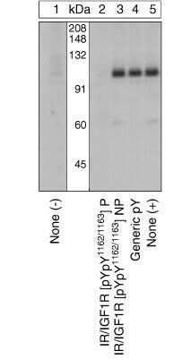
- Upregulation and Antibody-Peptide Competition. Extracts of CHO-T cells over-expressing the human insulin receptor unstimulated (1) or stimulated with 100 nM insulin for 10 min at 37°C (2-5) were resolved by SDS-PAGE on a 10% Tris-glycine gel and transferred to PVDF. The membrane was blocked with a 5% BSA-TBST buffer overnight at 4°C and incubated with the IR/IGF1R (pYpY1162/1163) antibody for two hours at room temperature in a 3% BSA-TBST buffer, following prior incubation with: no peptide (1, 5), the phosphopeptide immunogen (2), the non-phosphorylated peptide corresponding to the phosphopeptide immunogen (3), or a generic phosphotyrosine-containing peptide (4). After washing, the membrane was incubated with goat F (ab’)2 anti-rabbit IgG HRP-conjugate (Product # ALI4404) and signals were detected using the Pierce SuperSignal™method. The data show that only the phosphopeptide corresponding to IR/IGF1R (pYpY1162/1163) completely blocks the antibody signal, demonstrating the specificity of the antibody. The data also show the up-regulation of this site upon stimulation with insulin in this cell system.
Supportive validation
- Submitted by
- Invitrogen Antibodies (provider)
- Main image

- Experimental details
- Immunofluorescence analysis of IR/IGF1R (pYpY1162/1163) was done on 70% confluent log phase MCF7 cells treated with insulin (100nM for 5 min). The cells were fixed with 4% paraformaldehyde for 15 minutes, permeabilized with 0.25% Triton X-100 for 10 minutes, and blocked with 5% BSA for 1 hour at room temperature. The cells were labeled with IR/IGF1R (pYpY1162/1163) Rabbit polyclonal Antibody (Product # 44-804G) at 2 µg/mL in 1% BSA and incubated for 3 hours at room temperature and then labeled with Alexa Fluor 488 Goat Anti-Rabbit IgG Secondary Antibody (Product # A-11008) at a dilution of 1:400 for 30 minutes at room temperature (Panel a: green). Nuclei (Panel b: blue) were stained with SlowFade® Gold Antifade Mountant DAPI (Product # S36938). F-actin (Panel c: red) was stained with Alexa Fluor 594 Phalloidin (Product # A12381). Panel d is a merged image showing membrane localization. Panel e shows untreated MCF7 cells. Panel f shows no primary antibody control. The images were captured at 20X magnification.

Supportive validation
- Submitted by
- Invitrogen Antibodies (provider)
- Main image

- Experimental details
- Flow cytometry analysis of IR/IGF1R [pY1162/pY1163] was done on MCF7 cells treated with Insulin (100nM, 5 minutes). Cells were fixed with 70% ethanol for 10 minutes, permeabilized with 0.25% Tritonª X-100 for 20 minutes, and blocked with 5% BSA for 30 minutes at room temperature. Cells were labeled with IR/IGF1R [pY1162/pY1163] Rabbit Polyclonal Antibody (44804G, red histogram) or with rabbit isotype control (pink histogram) at 3-5 µg/million cells in 2.5% BSA. After incubation at room temperature for 2 hours, the cells were labeled with Alexa Fluor¨ 488 Goat Anti-Rabbit Secondary Antibody (A11008) at a dilution of 1:400 for 30 minutes at room temperature. The representative 10,000 cells were acquired and analyzed for each sample using an Attune¨ Acoustic Focusing Cytometer. The purple histogram represents unstained control cells and the green histogram represents no-primary-antibody control.

Supportive validation
- Submitted by
- Invitrogen Antibodies (provider)
- Main image

- Experimental details
- NULL

- Submitted by
- Invitrogen Antibodies (provider)
- Main image

- Experimental details
- NULL

- Submitted by
- Invitrogen Antibodies (provider)
- Main image

- Experimental details
- NULL
- Submitted by
- Invitrogen Antibodies (provider)
- Main image

- Experimental details
- NULL

- Submitted by
- Invitrogen Antibodies (provider)
- Main image

- Experimental details
- NULL

- Submitted by
- Invitrogen Antibodies (provider)
- Main image

- Experimental details
- Figure 4 Peripheral insulin signaling in 12-month-old mice on methionine restriction (MR) and control diet. Insulin signaling was assessed by administering either a saline ( n = 3) or low dose of insulin (0.8 mU g -1 ) ( n = 5-6) to mice, via i.p. injection, after a 5-h fast. Levels of phosphorylated IR (tyr 1162/1163), protein kinase B/Akt (ser473), S6 (ser235/236), IR-beta, total Akt, and total S6 were measured by immunoblotting in (A) epididymal white adipose tissue (WAT), (C) liver, and (E) gastrocnemius muscle in 12-month-old mice fed MR or control diet. Immunoblots were normalized to Ponceau S and total protein in (B) epididymal WAT, (D) liver, and (F) gastrocnemius muscle in 12-month-old mice fed MR or control diet. Data were analyzed as fold change relative to control-fed insulin-injected mice. Significance was calculated by two-tailed Student's t -test (* P < 0.05). Data are represented as mean +- SEM. Black hatch bars, 12-month-old control-fed mice injected with saline; black bars, 12-month-old control-fed mice injected with insulin; white crossed bars, 12-month-old MR-fed mice injected with saline; white bars, 12-month-old MR-fed mice injected with insulin.

- Submitted by
- Invitrogen Antibodies (provider)
- Main image

- Experimental details
- Figure 6 Effects of 48-h methionine restriction (MR) treatment on body weight, FGF21 and glucose homeostasis. (A) Body weight in mice fed MR or control diet ( n = 13). Significance was calculated by repeated measures two-way ANOVA with Bonferroni multiple comparison post hoc tests (* P < 0.05). (B) Serum FGF21 ( n = 13) and (C) hepatic gene expression of FGF21 ( n = 6) in mice fed MR or control diet. Data for gene expression were analyzed as fold change relative to control-fed mice. Significance was calculated by two-tailed Student's t -test (* P < 0.05). (D) Glucose tolerance as assessed by a glucose tolerance test (dose of glucose = 2 g kg -1 ) after a 5-h fast in mice fed MR or control diet ( n = 9-10). Significance was calculated by repeated measures two-way ANOVA with Bonferroni multiple comparison post hoc tests (* P < 0.05). Black bars/circles, control-fed mice; white bars/circles, MR-fed mice. Insulin signaling was assessed by administering either a saline ( n = 3) or high dose of insulin (10 mU g -1 ) ( n = 5-6) to mice, via i.p. injection, after a 5 h fast. (E) Levels of phosphorylated IR (tyr 1162/1163), protein kinase B/Akt (ser473), S6 (ser235/236), IR-beta, total Akt, and total S6 were measured by immunoblotting in liver in mice fed MR or control diet. (F) Immunoblots were normalized to Ponceau S and total protein in liver in mice fed MR or control diet. Data were analyzed as fold change relative to control-fed insulin-injected mice. Significance was calculated

- Submitted by
- Invitrogen Antibodies (provider)
- Main image

- Experimental details
- Figure 5 Compd. 23 increases liver cell insulin signaling, is orally bioavailable and reverses diabetes in obese mice (a-b) HepG2 hepatocytes were incubated overnight with 10 muM compound (Compd.) or dimethylsulfoxide (DMSO) and stimulated with 10 nM insulin for 5 min or left unstimulated (Unst.). Insulin receptor (IR) tyrosine phosphorylation was assessed by (a) Western blotting of anti-IR immunoprecipitations (representative of 2 independent experiments) and (b) phosphoIR (pIR) ELISA (data from 5 independent experiments, Compd. 23 : p=0.0079, Compd. 28 : p=0.1667). (c-g) Diet-induced obese (DIO) male B6 mice were treated with 0.05% w/w Compd. 23 in high-fat diet (HFD) or HFD alone for 2 weeks. (c) Body weight during treatment (HFD, n=17; Compd. 23 , n=13). (d) Intraperitoneal glucose tolerance test (IPGTT), p=0.0162 (HFD, n=8; Compd. 23 , n=6). (e) Fasting plasma insulin levels, relative to littermates fed HFD, p=0.0235 (HFD, n=4; Compd. 23 , n=4). (f-g) Mice were injected intraperitoneally with insulin and livers harvested after 10 min. (f) Liver IR tyrosine phosphorylation, p=0.0002 (HFD, n=7; Compd. 23 , n=6). (g) Western blot of liver homogenates (HFD, n=3; Compd. 23 , n=3). (h-i) DIO liver-specific LMPTP KO mice were treated with Compd. 23 (HFD, n=6; Compd. 23 , n=6) as in (c-g) . (h) IPGTT, p=0.6120. (i) Liver IR tyrosine phosphorylation, p=0.2791. (b-f,h-i) Mean+-SEM. *, p=0.05): (b) Kolmogorov-Smirnov test, (d,h) Two-Way ANOVA, (e) two-
