 Explore
 Validate
 Learn
Learn Western blot
Antibody data
- Antibody Data
- Antigen structure
- References [1]
- Comments [0]
- Validations
- Western blot [2]
- Immunocytochemistry [2]
- Immunohistochemistry [1]
Submit
Validation data
Reference
Comment
Report error
- Product number
- AF4654 - Provider product page

- Provider
- R&D Systems
- Product name
- Human/Mouse/Rat CDK2 Antibody
- Antibody type
- Polyclonal
- Description
- Antigen Affinity-purified. Detects human, mouse, and rat CDK2 in Western blots. In Western blots, less than 1% cross-reactivity with recombinant human (rh) CDK4 and rhCDK6 is observed.
- Reactivity
- Human, Mouse, Rat
- Host
- Goat
- Conjugate
- Unconjugated
- Antigen sequence
P24941- Isotype
- IgG
- Vial size
- 100 ug
- Concentration
- LYOPH
- Storage
- Use a manual defrost freezer and avoid repeated freeze-thaw cycles. 12 months from date of receipt, -20 to -70 °C as supplied. 1 month, 2 to 8 °C under sterile conditions after reconstitution. 6 months, -20 to -70 °C under sterile conditions after reconstitution.
Submitted references Chemotherapy-induced differential cell cycle arrest in B-cell lymphomas affects their sensitivity to Wee1 inhibition.
Wang X, Chen Z, Mishra AK, Silva A, Ren W, Pan Z, Wang JH
Haematologica 2018 Mar;103(3):466-476
Haematologica 2018 Mar;103(3):466-476
No comments: Submit comment
Supportive validation
- Submitted by
- R&D Systems (provider)
- Main image

- Experimental details
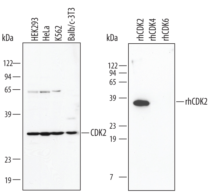
- Detection of Human/Mouse CDK2 by Western Blot. Western blot shows lysates of HEK293 human embryonic kidney cell line, HeLa human cervical epithelial carcinoma cell line, K562 human chronic myelogenous leukemia cell line, and Balb/3T3 mouse embryonic fibroblast cell line. PVDF membrane was probed with 1 µg/mL Goat Anti-Human/Mouse CDK2 Antigen Affinity-purified Polyclonal Antibody (Catalog # AF4654) followed by HRP-conjugated Anti-Goat IgG Secondary Antibody (Catalog # HAF017). For additional reference, recombinant human CDK2, CDK4, and CDK6 (5 ng/lane) were included. A specific band for CDK2 was detected at approximately 34 kDa (as indicated). This experiment was conducted under reducing conditions and using Immunoblot Buffer Group 1.
- Submitted by
- R&D Systems (provider)
- Main image

- Experimental details
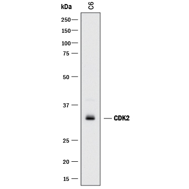
- Detection of Rat CDK2 by Western Blot. Western blot shows lysates of C6 rat glioma cell line. PVDF membrane was probed with 1 µg/mL of Goat Anti-Human/Mouse CDK2 Antigen Affinity-purified Polyclonal Antibody (Catalog # AF4654) followed by HRP-conjugated Anti-Goat IgG Secondary Antibody (Catalog # HAF017). A specific band was detected for CDK2 at approximately 34 kDa (as indicated). This experiment was conducted under reducing conditions and using Immunoblot Buffer Group 1.
Supportive validation
- Submitted by
- R&D Systems (provider)
- Main image

- Experimental details
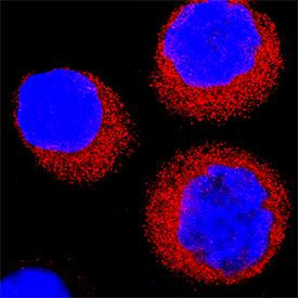
- CDK2 in 3T3-L1 Mouse Cell Line. CDK2 was detected in immersion fixed 3T3-L1 mouse embryonic fibroblast adipose-like cell line using Goat Anti-Human/Mouse CDK2 Antigen Affinity-purified Polyclonal Antibody (Catalog # AF4654) at 15 µg/mL for 3 hours at room temperature. Cells were stained using the NorthernLights™ 557-conjugated Anti-Goat IgG Secondary Antibody (red; Catalog # NL001) and counterstained with DAPI (blue). Specific staining was localized to cytoplasm and nuclei. View our protocol for Fluorescent ICC Staining of Cells on Coverslips.

- Submitted by
- R&D Systems (provider)
- Main image

- Experimental details
- CDK2 in K562 Human Cell Line. CDK2 was detected in immersion fixed K562 human chronic myelogenous leukemia cell line using Goat Anti-Human/Mouse CDK2 Antigen Affinity-purified Polyclonal Antibody (Catalog # AF4654) at 5 µg/mL for 3 hours at room temperature. Cells were stained using the NorthernLights™ 557-conjugated Anti-Goat IgG Secondary Antibody (red; Catalog # NL001) and counterstained with DAPI (blue). Specific staining was localized to cytoplasm. View our protocol for Fluorescent ICC Staining of Non-adherent Cells.
Supportive validation
- Submitted by
- R&D Systems (provider)
- Main image
- Experimental details
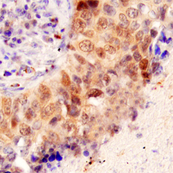
- CDK2 in Human Lung Cancer Tissue. CDK2 was detected in immersion fixed paraffin-embedded sections of human lung cancer tissue using Goat Anti-Human/Mouse CDK2 Antigen Affinity-purified Polyclonal Antibody (Catalog # AF4654) at 3 µg/mL for 1 hour at room temperature followed by incubation with the Anti-Goat IgG VisUCyte™ HRP Polymer Antibody (Catalog # VC004). Tissue was stained using DAB (brown) and counterstained with hematoxylin (blue). Specific staining was localized to cytoplasm and cancer cell nuclei. View our protocol for IHC Staining with VisUCyte HRP Polymer Detection Reagents.
