 Explore
 Validate
 Learn
Learn Western blot
Antibody data
- Antibody Data
- Antigen structure
- References [2]
- Comments [0]
- Validations
- Western blot [2]
- Immunohistochemistry [3]
Submit
Validation data
Reference
Comment
Report error
- Product number
- AF3389 - Provider product page

- Provider
- R&D Systems
- Product name
- Human/Mouse/Rat Src Antibody
- Antibody type
- Polyclonal
- Description
- Antigen Affinity-purified. Detects endogenous human, mouse, and rat Src in Western blots.
- Reactivity
- Human, Mouse, Rat
- Host
- Goat
- Conjugate
- Unconjugated
- Antigen sequence
P12931- Isotype
- IgG
- Vial size
- 100 ug
- Concentration
- LYOPH
- Storage
- Use a manual defrost freezer and avoid repeated freeze-thaw cycles. 12 months from date of receipt, -20 to -70 °C as supplied. 1 month, 2 to 8 °C under sterile conditions after reconstitution. 6 months, -20 to -70 °C under sterile conditions after reconstitution.
Submitted references An IL13Rα2 peptide exhibits therapeutic activity against metastatic colorectal cancer.
Clonorchis sinensis excretory-secretory products promote the migration and invasion of cholangiocarcinoma cells by activating the integrin β4-FAK/Src signaling pathway.
Bartolomé RA, Jaén M, Casal JI
British journal of cancer 2018 Oct;119(8):940-949
British journal of cancer 2018 Oct;119(8):940-949
Clonorchis sinensis excretory-secretory products promote the migration and invasion of cholangiocarcinoma cells by activating the integrin β4-FAK/Src signaling pathway.
Pak JH, Bashir Q, Kim IK, Hong SJ, Maeng S, Bahk YY, Kim TS
Molecular and biochemical parasitology 2017 Jun;214:1-9
Molecular and biochemical parasitology 2017 Jun;214:1-9
No comments: Submit comment
Supportive validation
- Submitted by
- R&D Systems (provider)
- Main image

- Experimental details
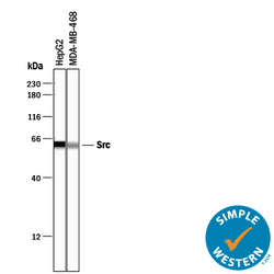
- Detection of Human and Rat Src by Western Blot. Western blot shows lysates of HepG2 human hepatocellular carcinoma cell line, MBA-MB-468 human breast cancer cell line, and Rat-2 rat embryonic fibroblast cell line. PVDF membrane was probed with 0.5 µg/mL of Human/Mouse/Rat Src Antigen Affinity-purified Polyclonal Antibody (Catalog # AF3389) followed by HRP-conjugated Anti-Goat IgG Secondary Antibody (Catalog # HAF109). A specific band was detected for Src at approximately 60 kDa (as indicated). This experiment was conducted under reducing conditions and using Immunoblot Buffer Group 1.

- Submitted by
- R&D Systems (provider)
- Main image
- Experimental details
- Detection of Human Src by Simple WesternTM. Simple Western lane view shows lysates of HepG2 human hepatocellular carcinoma cell line and MDA-MB-468 human breast cancer cell line, loaded at 0.2 mg/mL. A specific band was detected for Src at approximately 62 kDa (as indicated) using 25 µg/mL of Goat Anti-Human/Mouse/Rat Src Antigen Affinity-purified Polyclonal Antibody (Catalog # AF3389) followed by 1:50 dilution of HRP-conjugated Anti-Goat IgG Secondary Antibody (Catalog # HAF109). This experiment was conducted under reducing conditions and using the 12-230 kDa separation system.

Supportive validation
- Submitted by
- R&D Systems (provider)
- Main image

- Experimental details
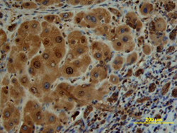
- Src in Human Liver Cancer Tissue. Src was detected in immersion fixed paraffin-embedded sections of human liver cancer tissue using Human/Mouse/Rat Src Antigen Affinity-purified Polyclonal Antibody (Catalog # AF3389) at 15 µg/mL overnight at 4 °C. Tissue was stained using the Anti-Goat HRP-DAB Cell & Tissue Staining Kit (brown; Catalog # CTS008) and counterstained with hematoxylin (blue). View our protocol for Chromogenic IHC Staining of Paraffin-embedded Tissue Sections.

- Submitted by
- R&D Systems (provider)
- Main image
- Experimental details
- Src in Human Liver. Src was detected in immersion fixed paraffin-embedded sections of human liver array using Human/Mouse/Rat Src Antigen Affinity-purified Polyclonal Antibody (Catalog # AF3389) at 10 µg/mL overnight at 4 °C. Tissue was stained using the Anti-Goat HRP-DAB Cell & Tissue Staining Kit (brown; Catalog # CTS008) and counterstained with hematoxylin (blue). View our protocol for Chromogenic IHC Staining of Paraffin-embedded Tissue Sections.

- Submitted by
- R&D Systems (provider)
- Main image
- Experimental details
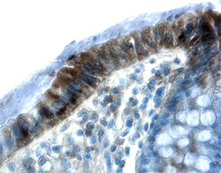
- Src in Human Colon. Src was detected in immersion fixed paraffin-embedded sections of human colon using 15 µg/mL Human/Mouse/Rat Src Antigen Affinity-purified Polyclonal Antibody (Catalog # AF3389) overnight at 4 °C. Tissue was stained with the Anti-Goat HRP-DAB Cell & Tissue Staining Kit (brown; Catalog # CTS008) and counterstained with hematoxylin (blue). View our protocol for Chromogenic IHC Staining of Paraffin-embedded Tissue Sections.
