 Explore
 Validate
 Learn
Learn Western blot
Antibody data
- Antibody Data
- Antigen structure
- References [1]
- Comments [0]
- Validations
- Western blot [2]
- Immunohistochemistry [1]
Submit
Validation data
Reference
Comment
Report error
- Product number
- AF3918 - Provider product page

- Provider
- Novus Biologicals
- Product name
- Rabbit Polyclonal Ribosomal Protein S6/RPS6 Antibody
- Antibody type
- Polyclonal
- Description
- Immunogen affinity purified. Detects human, mouse, and rat ribosomal protein S6 when dually phosphorylated at S235 and S236.
- Reactivity
- Human, Mouse, Rat
- Host
- Rabbit
- Conjugate
- Unconjugated
- Isotype
- IgG
- Vial size
- 100 ug
- Concentration
- LYOPH
- Storage
- Use a manual defrost freezer and avoid repeated freeze-thaw cycles. 12 months from date of receipt, -20 to -70 degreesC as supplied. 1 month, 2 to 8 degreesC under sterile conditions after reconstitution. 6 months, -20 to -70 degreesC under sterile conditions after reconstitution.
Submitted references The protein-tyrosine phosphatase, SRC homology-2 domain containing protein tyrosine phosphatase-2, is a crucial mediator of exogenous insulin-like growth factor signaling to human trophoblast.
Forbes K, West G, Garside R, Aplin JD, Westwood M
Endocrinology 2009 Oct;150(10):4744-54
Endocrinology 2009 Oct;150(10):4744-54
No comments: Submit comment
Supportive validation
- Submitted by
- Novus Biologicals (provider)
- Main image
- Experimental details
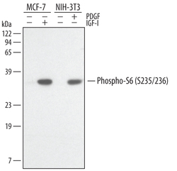
- Detection of Human and Mouse Phospho-Ribosomal Protein S6 (S235/S236) by Western Blot. Western blot shows lysates of MCF-7 human breast cancer cell line untreated (-) or treated (+) with 100 ng/mL Recombinant Human IGF-1 (Catalog # 291-G1) for 20 minutes and NIH-3T3 mouse embryonic fibroblast cell line untreated or treated with 10 ng/mL Human PDGF (Catalog # 120-HD) for 20 minutes. PVDF membrane was probed with 0.5 µg/mL of Rabbit Anti-Human/Mouse/Rat Phospho-Ribosomal Protein S6 (S235/S236) Antigen Affinity-purified Polyclonal Antibody (Catalog # AF3918), followed by HRP-conjugated Anti-Rabbit IgG Secondary Antibody (Catalog # HAF008). A specific band was detected for Phospho-Ribosomal Protein S6 (S235/S236) at approximately 32 kDa (as indicated). This experiment was conducted under reducing conditions and using Immunoblot Buffer Group 1.
- Submitted by
- Novus Biologicals (provider)
- Main image

- Experimental details
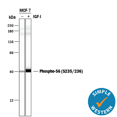
- Detection of Human Phospho-Ribosomal Protein S6/RPS6 (S235/S236) by Simple WesternTM. Simple Western lane view shows lysates of MCF-7 human breast cancer cell line untreated (-) or treated (+) with 100 ng/mL Recombinant Human IGF-I (Catalog # 291-G1) for 20 minutes, loaded at 0.5 mg/mL. A specific band was detected for Phospho-Ribosomal Protein S6/RPS6 (S235/S236) at approximately 40 kDa (as indicated) using 5 µg/mL of Rabbit Anti-Human/Mouse/Rat Phospho-Ribosomal Protein S6/RPS6 (S235/S236) Antigen Affinity-purified Polyclonal Antibody (Catalog # AF3918). This experiment was conducted under reducing conditions and using the 12-230 kDa separation system.
Supportive validation
- Submitted by
- Novus Biologicals (provider)
- Main image
- Experimental details
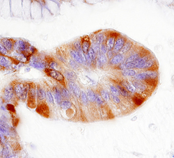
- Ribosomal Protein S6 in Human Colon Cancer Tissue. Ribosomal Protein S6 was detected in immersion fixed paraffin-embedded sections of human colon cancer tissue using 5 µg/mL Rabbit Anti-Human/Mouse/Rat Phospho-Ribosomal Protein S6 (S235/S236) Antigen Affinity-purified Polyclonal Antibody (Catalog # AF3918) overnight at 4 °C. Tissue was stained with the Anti-Rabbit HRP-DAB Cell & Tissue Staining Kit (brown; Catalog # CTS005) and counterstained with hematoxylin (blue). Specific labeling was localized to vascular endothelial cell within glomeruli. View our protocol for Chromogenic IHC Staining of Paraffin-embedded Tissue Sections.
