 Explore
 Validate
 Learn
LearnAER-031-200UL
antibody from Invitrogen Antibodies
Targeting: EFNA1
ECKLG, EPLG1, GMAN, LERK1, TNFAIP4
 Western blot
Antibody data
- Antibody Data
- Antigen structure
- References [0]
- Comments [0]
- Validations
- Western blot [4]
- Immunocytochemistry [1]
- Flow cytometry [1]
Submit
Validation data
Reference
Comment
Report error
- Product number
- AER-031-200UL - Provider product page

- Provider
- Invitrogen Antibodies
- Product name
- Ephrin-A1 (extracellular) Polyclonal Antibody
- Antibody type
- Polyclonal
- Antigen
- Other
- Reactivity
- Human, Mouse, Rat
- Host
- Rabbit
- Isotype
- IgG
- Vial size
- 200 µL
- Concentration
- 0.8 mg/mL
- Storage
- -20° C, Avoid Freeze/Thaw Cycles
No comments: Submit comment
Supportive validation
- Submitted by
- Invitrogen Antibodies (provider)
- Main image

- Experimental details
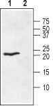
- Western blot analysis of rat heart membranes: - 1. Anti-Ephrin-A1 (extracellular) Antibody (#AER-031), (1:400). 2. Anti-Ephrin-A1 (extracellular) Antibody , preincubated with Ephrin-A1 (extracellular) Blocking Peptide (#BLP-ER031).
- Submitted by
- Invitrogen Antibodies (provider)
- Main image

- Experimental details
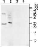
- Western blot analysis of mouse brain membranes (lanes 1 and 3) and human Malme-3M melanoma cell lysate (lanes 2 and 4): - 1,2. Anti-Ephrin-A1 (extracellular) Antibody (#AER-031), (1:200).3,4. Anti-Ephrin-A1 (extracellular) Antibody , preincubated with Ephrin-A1 (extracellular) Blocking Peptide (#BLP-ER031).
- Submitted by
- Invitrogen Antibodies (provider)
- Main image

- Experimental details
- Western blot analysis of rat heart membranes: - 1. Anti-Ephrin-A1 (extracellular) Antibody (#AER-031), (1:400). 2. Anti-Ephrin-A1 (extracellular) Antibody , preincubated with Ephrin-A1 (extracellular) Blocking Peptide (#BLP-ER031).

- Submitted by
- Invitrogen Antibodies (provider)
- Main image

- Experimental details
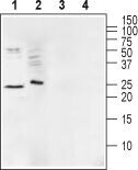
- Western blot analysis of mouse brain membranes (lanes 1 and 3) and human Malme-3M melanoma cell lysate (lanes 2 and 4): - 1,2. Anti-Ephrin-A1 (extracellular) Antibody (#AER-031), (1:200).3,4. Anti-Ephrin-A1 (extracellular) Antibody , preincubated with Ephrin-A1 (extracellular) Blocking Peptide (#BLP-ER031).
Supportive validation
- Submitted by
- Invitrogen Antibodies (provider)
- Main image
- Experimental details
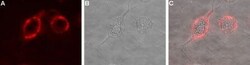
- Expression of Ephrin-A1 in rat PC12 cells - Cell surface detection of Ephrin-A1 in live intact rat PC12 pheochromocytoma cells. A. Extracellular staining of cells with Anti-Ephrin-A1 (extracellular) Antibody (#AER-031), (1:50), followed by goat Anti-rabbit-AlexaFluor-594 secondary Antibody (red). B. Live view of the cells. C. Merge of A and B.

Supportive validation
- Submitted by
- Invitrogen Antibodies (provider)
- Main image

- Experimental details
- Cell surface detection ofEphrin-A1by indirect flow cytometry in live intacthumanJurkatT-cellleukemiacells: - (black line) cells. (red) Cells+ goat- Anti-rabbit-FITC. (green) Cells + Anti-Ephrin-A1 (extracellular) Antibody (#AER-031), (5μg) + goat- Anti-rabbit-FITC.
