Explore
 Validate
 Learn
Learn Western blot
 Immunocytochemistry
Antibody data
- Antibody Data
- Antigen structure
- References [2]
- Comments [0]
- Validations
- Western blot [1]
- Immunocytochemistry [1]
- Immunohistochemistry [8]
Submit
Validation data
Reference
Comment
Report error
- Product number
- HPA021616 - Provider product page

- Provider
- Atlas Antibodies
- Proper citation
- Atlas Antibodies Cat#HPA021616, RRID:AB_1848342
- Product name
- Anti-EZR
- Antibody type
- Polyclonal
- Reactivity
- Human, Mouse, Rat
- Host
- Rabbit
- Conjugate
- Unconjugated
- Antigen sequence
REEKHQKQLERQQLETEKKRRETVEREKEQMMREK
EELMLRLQDYEEKTKKAERELSEQIQRALQLEEER
KRAQEEAERLEADRMAALRAKEELERQAVDQIKSQ
EQLAAELAEYTAKIALLE- Isotype
- IgG
- Vial size
- 100 µl
- Storage
- Store at +4°C for short term storage. Long time storage is recommended at -20°C.
Submitted references Incident urothelial cancer in the Malmö Diet and Cancer Study: cohort characteristics and further validation of ezrin as a prognostic biomarker.
Reduced expression of ezrin in urothelial bladder cancer signifies more advanced tumours and an impaired survival: validatory study of two independent patient cohorts.
Wennersten C, Andersson G, Boman K, Nodin B, Gaber A, Jirström K
Diagnostic pathology 2014 Oct 3;9:189
Diagnostic pathology 2014 Oct 3;9:189
Reduced expression of ezrin in urothelial bladder cancer signifies more advanced tumours and an impaired survival: validatory study of two independent patient cohorts.
Andersson G, Wennersten C, Gaber A, Boman K, Nodin B, Uhlén M, Segersten U, Malmström PU, Jirström K
BMC urology 2014 May 12;14:36
BMC urology 2014 May 12;14:36
No comments: Submit comment
Enhanced validation
- Submitted by
- Atlas Antibodies (provider)
- Enhanced method
- Genetic validation
- Main image
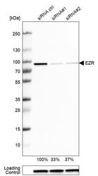
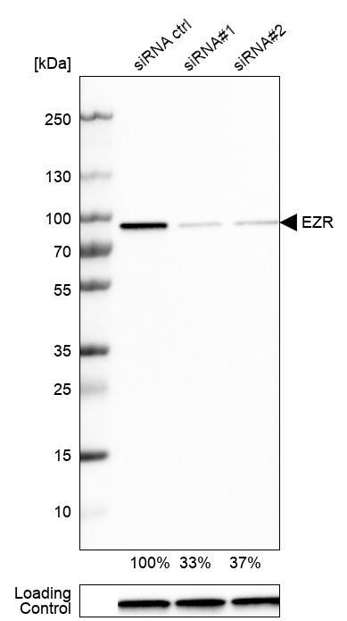
- Experimental details
- Western blot analysis in U-251MG cells transfected with control siRNA, target specific siRNA probe #1 and #2, using Anti-EZR antibody. Remaining relative intensity is presented. Loading control: Anti-GAPDH.
Supportive validation
- Submitted by
- Atlas Antibodies (provider)
- Main image
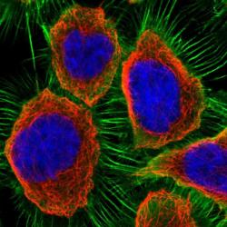
- Experimental details
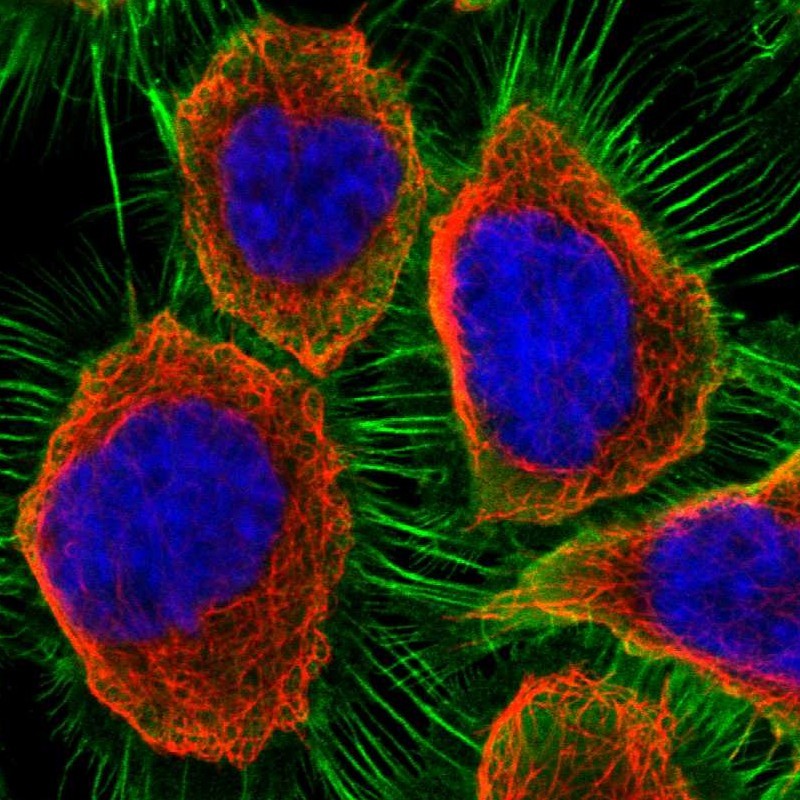
- Immunofluorescent staining of human cell line A-431 shows localization to plasma membrane.
- Sample type
- HUMAN
Enhanced validation
Supportive validation
- Submitted by
- Atlas Antibodies (provider)
- Enhanced method
- Orthogonal validation
- Main image
- Experimental details
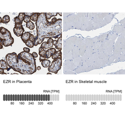
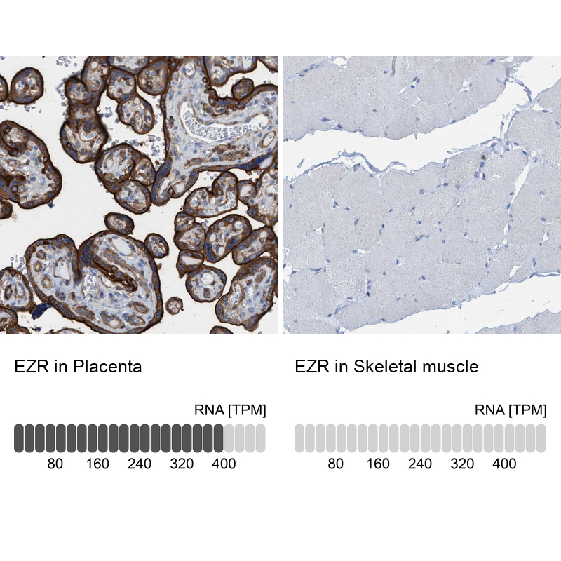
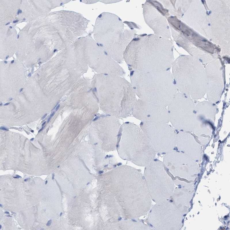
- Immunohistochemistry analysis in human placenta and skeletal muscle tissues using HPA021616 antibody. Corresponding EZR RNA-seq data are presented for the same tissues.
- Sample type
- HUMAN
Supportive validation
- Submitted by
- Atlas Antibodies (provider)
- Main image

- Experimental details
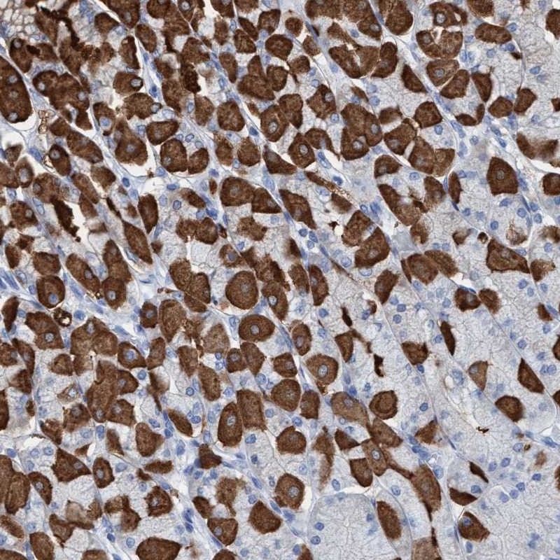
- Immunohistochemical staining of human stomach (upper) shows strong cytoplasmic positivity in parietal cells.
- Submitted by
- Atlas Antibodies (provider)
- Main image
- Experimental details
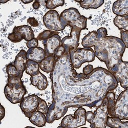
- Immunohistochemical staining of human placenta shows high expression.
- Sample type
- HUMAN

- Submitted by
- Atlas Antibodies (provider)
- Main image
- Experimental details
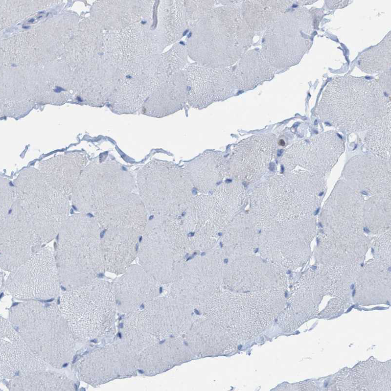
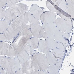
- Immunohistochemical staining of human skeletal muscle shows low expression as expected.
- Sample type
- HUMAN
- Submitted by
- Atlas Antibodies (provider)
- Main image
- Experimental details
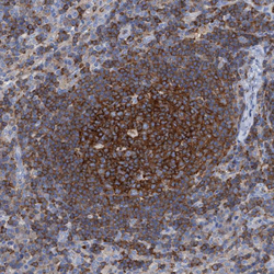
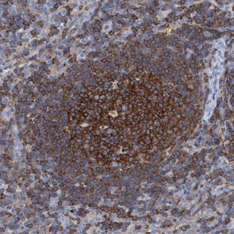
- Immunohistochemical staining of human lymph node shows strong membranous positivity in germinal center cells.
- Sample type
- HUMAN
- Submitted by
- Atlas Antibodies (provider)
- Main image
- Experimental details
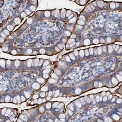
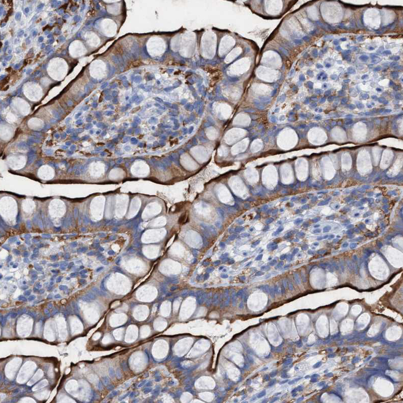
- Immunohistochemical staining of human small intestine shows strong positivity in apical membrane in glandular cells.
- Sample type
- HUMAN
- Submitted by
- Atlas Antibodies (provider)
- Main image
- Experimental details
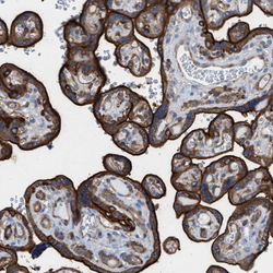
- Immunohistochemical staining of human placenta shows strong membranous positivity in trophoblastic cells.
- Sample type
- HUMAN

- Submitted by
- Atlas Antibodies (provider)
- Main image
- Experimental details
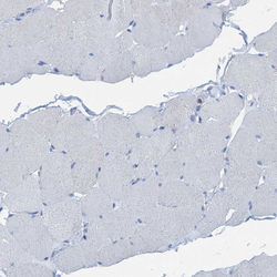
- Immunohistochemical staining of human skeletal muscle shows no positivity in myocytes as expected.
- Sample type
- HUMAN