Explore
 Validate
 Learn
Learn Western blot
Antibody data
- Antibody Data
- Antigen structure
- References [2]
- Comments [0]
- Validations
- Western blot [7]
- Immunocytochemistry [2]
- Immunohistochemistry [1]
Submit
Validation data
Reference
Comment
Report error
- Product number
- PA5-22309 - Provider product page

- Provider
- Invitrogen Antibodies
- Product name
- NDUFS1 Polyclonal Antibody
- Antibody type
- Polyclonal
- Antigen
- Recombinant protein fragment
- Description
- Recommended positive controls: Molt-4, mouse brain, rat brain. Predicted reactivity: Mouse (90%), Rat (93%), Zebrafish (82%), Xenopus laevis (85%), Chicken (86%), Chimpanzee (100%), Bovine (96%). Store product as a concentrated solution. Centrifuge briefly prior to opening the vial.
- Reactivity
- Human, Mouse, Rat
- Host
- Rabbit
- Isotype
- IgG
- Vial size
- 100 µL
- Concentration
- 0.57 mg/mL
- Storage
- Store at 4°C short term. For long term storage, store at -20°C, avoiding freeze/thaw cycles.
Submitted references CRISPR-Cas9 screen identifies oxidative phosphorylation as essential for cancer cell survival at low extracellular pH.
CCN6 regulates mitochondrial function.
Michl J, Wang Y, Monterisi S, Blaszczak W, Beveridge R, Bridges EM, Koth J, Bodmer WF, Swietach P
Cell reports 2022 Mar 8;38(10):110493
Cell reports 2022 Mar 8;38(10):110493
CCN6 regulates mitochondrial function.
Patra M, Mahata SK, Padhan DK, Sen M
Journal of cell science 2016 Jul 15;129(14):2841-51
Journal of cell science 2016 Jul 15;129(14):2841-51
No comments: Submit comment
Supportive validation
- Submitted by
- Invitrogen Antibodies (provider)
- Main image
- Experimental details
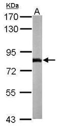
- Western blot analysis of NDUFS1 using 50 µg of mouse brain lysate. Samples were loaded onto a 7.5% SDS-PAGE gel and probed with a NDUFS1 polyclonal antibody (Product # PA5-22309) at a dilution of 1:1000.

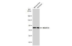
- Submitted by
- Invitrogen Antibodies (provider)
- Main image

- Experimental details
- Western blot analysis of NDUFS1 using 50 µg of rat brain lysate. Samples were loaded onto a 12% SDS-PAGE gel and probed with a NDUFS1 polyclonal antibody (Product # PA5-22309) at a dilution of 1:1000.

- Submitted by
- Invitrogen Antibodies (provider)
- Main image
- Experimental details
- Western blot analysis of NDUFS1 using 30 µg of MOLT4 lysate. Samples were loaded onto a 12% SDS-PAGE gel and probed with a NDUFS1 polyclonal antibody (Product # PA5-22309) at a dilution of 1:1000.

- Submitted by
- Invitrogen Antibodies (provider)
- Main image
- Experimental details
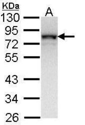
- Western Blot using NDUFS1 Polyclonal Antibody (Product # PA5-22309). HepG2 and mitochondria extracts (30 µg) were separated by SDS-PAGE, and the membrane was blotted with NDUFS1 Polyclonal Antibody (Product # PA5-22309) diluted at 1:1,000. The HRP-conjugated anti-rabbit IgG antibody was used to detect the primary antibody.

- Submitted by
- Invitrogen Antibodies (provider)
- Main image
- Experimental details
- Western Blot using NDUFS1 Polyclonal Antibody (Product # PA5-22309). Various tissue extracts (50 µg) were separated by 7.5% SDS-PAGE, and the membrane was blotted with NDUFS1 Polyclonal Antibody (Product # PA5-22309) diluted at 1:1,000. The HRP-conjugated anti-rabbit IgG antibody was used to detect the primary antibody.

- Submitted by
- Invitrogen Antibodies (provider)
- Main image
- Experimental details
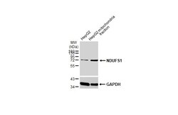
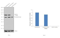
- Knockdown of NDUFS1 was achieved by transfecting HeLa cells with NDUFS1 specific siRNAs (Silencer® select Product # s9382, s9383). Western blot analysis (Fig. a) was performed using whole cell extracts from the NDUFS1 knockdown cells (lane 3), non-specific scrambled siRNA transfected cells (lane 2) and untransfected cells (lane 1). The blots were probed with NDUFS1 Polyclonal Antibody (Product # PA5-22309, 1:1000 dilution) and Goat anti-Rabbit IgG (H+L) Superclonal™ Secondary Antibody, HRP conjugate (Product # A27036, 0.25 µg/ml, 1:4000 dilution). Densitometric analysis of this western blot is shown in histogram (Fig. b). Decrease in signal upon siRNA mediated knock down confirms that antibody is specific to NDUFS1.

- Submitted by
- Invitrogen Antibodies (provider)
- Main image
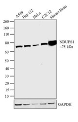
- Experimental details
- Western blot analysis was performed on membrane enriched extracts (30 µg lysate) of A549 (Lane 1), Hep G2 (Lane 2), HeLa (Lane 3), C2C12 (Lane 4) and tissue extract of Mouse Brain (Lane 5). The blot was probed with Anti- NDUFS1 Polyclonal Antibody (Product # PA5-22309, 1µg/ml) and detected by chemiluminescence using Goat anti Rabbit IgG (H+L) Superclonal™ Secondary Antibody, HRP conjugate (Product # A27036, 0.25 µg/ml, 1:4000 dilution). A 75 kDa band corresponding to NDUFS1 was detected across the cell lines and tissue tested.

Supportive validation
- Submitted by
- Invitrogen Antibodies (provider)
- Main image

- Experimental details
- Immunofluorescent analysis of NDUFS1 in methanol-fixed A431 cells using a NDUFS1 polyclonal antibody (Product # PA5-22309) at a 1:200 dilution.

- Submitted by
- Invitrogen Antibodies (provider)
- Main image

- Experimental details
- Immunofluorescence analysis of NDUFS1 was performed using 70% confluent log phase HeLa cells. The cells were fixed with 4% paraformaldehyde for 10 minutes, permeabilized with 0.1% Triton™ X-100 for 10 minutes, and blocked with 1% BSA for 1 hour at room temperature. The cells were labeled with NDUFS1 Rabbit Polyclonal Antibody (Product # PA5-22309) at 5 µg/mL in 0.1% BSA and incubated overnight at 4 degree and then labeled with Goat anti-Rabbit IgG (H+L) Superclonal™ Secondary Antibody, Alexa Fluor® 488 conjugate (Product # A27034) at a dilution of 1:2000 for 45 minutes at room temperature (Panel a: green). Nuclei (Panel b: blue) were stained with SlowFade® Gold Antifade Mountant with DAPI (Product # S36938). Mitochondria (Panel c: red) was stained with Mitotracker Red CMXRos (Product # M7512). Panel d represents the merged image showing mitochondrial localization. Panel e represents control cells with no primary antibody to assess background. The images were captured at 60X magnification.

Supportive validation
- Submitted by
- Invitrogen Antibodies (provider)
- Main image

- Experimental details
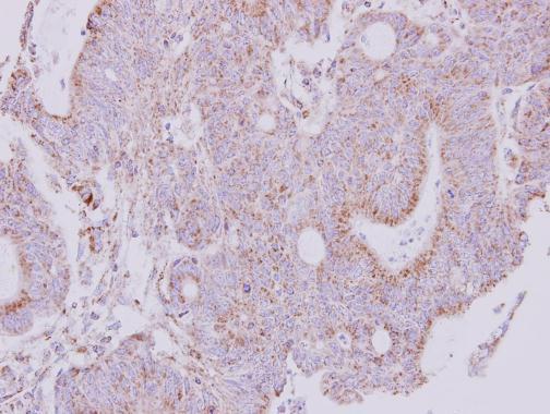
- Immunohistochemical analysis of paraffin-embedded human colon carcinoma, using NDUFS1 (Product # PA5-22309) antibody at 1:250 dilution. Antigen Retrieval: EDTA based buffer, pH 8.0, 15 min.