 Explore
 Validate
 Learn
Learn Western blot
Antibody data
- Antibody Data
- Antigen structure
- References [3]
- Comments [0]
- Validations
- Western blot [1]
- Immunocytochemistry [1]
- Other assay [1]
Submit
Validation data
Reference
Comment
Report error
- Product number
- 44-1000G - Provider product page

- Provider
- Invitrogen Antibodies
- Product name
- Phospho-PDGFRA/PDGFRB (Tyr572, Tyr574) Polyclonal Antibody
- Antibody type
- Polyclonal
- Antigen
- Synthetic peptide
- Reactivity
- Human, Mouse
- Host
- Rabbit
- Isotype
- IgG
- Vial size
- 100 µL
- Storage
- -20°C
Submitted references Lycorine inhibits angiogenesis by docking to PDGFRα.
Hepatic Stellate Cell-Specific Platelet-Derived Growth Factor Receptor-α Loss Reduces Fibrosis and Promotes Repair after Hepatocellular Injury.
Platelet-Derived Growth Factor Receptor α Contributes to Human Hepatic Stellate Cell Proliferation and Migration.
Lv F, Li X, Wang Y
BMC cancer 2022 Aug 10;22(1):873
BMC cancer 2022 Aug 10;22(1):873
Hepatic Stellate Cell-Specific Platelet-Derived Growth Factor Receptor-α Loss Reduces Fibrosis and Promotes Repair after Hepatocellular Injury.
Kikuchi A, Singh S, Poddar M, Nakao T, Schmidt HM, Gayden JD, Sato T, Arteel GE, Monga SP
The American journal of pathology 2020 Oct;190(10):2080-2094
The American journal of pathology 2020 Oct;190(10):2080-2094
Platelet-Derived Growth Factor Receptor α Contributes to Human Hepatic Stellate Cell Proliferation and Migration.
Kikuchi A, Pradhan-Sundd T, Singh S, Nagarajan S, Loizos N, Monga SP
The American journal of pathology 2017 Oct;187(10):2273-2287
The American journal of pathology 2017 Oct;187(10):2273-2287
No comments: Submit comment
Supportive validation
- Submitted by
- Invitrogen Antibodies (provider)
- Main image
- Experimental details
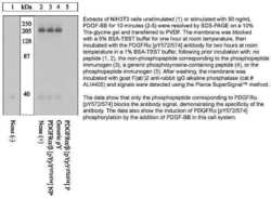
- Western blot analysis of Phospho-PDGFRA/PDGFRB (Tyr572, Tyr574) using a polyclonal antibody (Product # 44-1000G).

Supportive validation
- Submitted by
- Invitrogen Antibodies (provider)
- Main image

- Experimental details
- Immunofluorescence analysis of PDGFR alpha + beta (pYpY572/574) was done on 70% confluent log phase MCF7 cells treated with PDGF (50 ng/mL for 10 min). The cells were fixed with 4% paraformaldehyde for 15 minutes, permeabilized with 0.25% Triton X-100 for 10 minutes, and blocked with 5% BSA for 1 hour at room temperature. The cells were labeled with PDGFR alpha + beta (pYpY572/574) Rabbit polyclonal Antibody (Product # 44-1000G) at 2 µg/mL in 1% BSA and incubated for 3 hours at room temperature and then labeled with Alexa Fluor 488 Goat Anti-Rabbit IgG Secondary Antibody (Product # A-11008) at a dilution of 1:400 for 30 minutes at room temperature (Panel a: green). Nuclei (Panel b: blue) were stained with SlowFade® Gold Antifade Mountant DAPI (Product # S36938). F-actin (Panel c: red) was stained with Alexa Fluor 594 Phalloidin (Product # A12381). Panel d is a merged image showing cytoplasmic and membrane localization. Panel e shows untreated cells. Panel f shows no primary antibody control. The images were captured at 20X magnification.

Supportive validation
- Submitted by
- Invitrogen Antibodies (provider)
- Main image

- Experimental details
- NULL
