Explore
 Validate
 Learn
Learn Immunocytochemistry
 Immunohistochemistry
Antibody data
- Antibody Data
- Antigen structure
- References [0]
- Comments [0]
- Validations
- Immunocytochemistry [1]
- Immunohistochemistry [6]
Submit
Validation data
Reference
Comment
Report error
- Product number
- AMAb91501 - Provider product page

- Provider
- Atlas Antibodies
- Product name
- Anti-HDAC5
- Antibody type
- Monoclonal
- Antigen
- Synthetic peptide
- Reactivity
- Human
- Host
- Mouse
- Conjugate
- Unconjugated
- Antigen sequence
KSKEPTPGGLNHSLPQHPK- Epitope
- Binds to an epitope located within the peptide sequence - as determined by overlapping synthetic peptides.
- Isotype
- IgG
- Antibody clone number
- CL8121
- Vial size
- 100 µl
- Storage
- Store at +4°C for short term storage. Long time storage is recommended at -20°C.
No comments: Submit comment
Supportive validation
- Submitted by
- Atlas Antibodies (provider)
- Main image
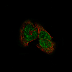
- Experimental details
- Immunofluorescence staining of RH-30 cells using the Anti-HDAC5 monoclonal antibody, showing specific staining in the nucleoplasm and cytosol in green. Microtubule- and nuclear probes are visualized in red and blue, respectively (where available).
- Sample type
- HUMAN

Enhanced validation
Supportive validation
- Submitted by
- Atlas Antibodies (provider)
- Enhanced method
- Orthogonal validation
- Main image
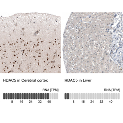
- Experimental details
- Immunohistochemistry analysis in human cerebral cortex and liver tissues using AMAb91501 antibody. Corresponding HDAC5 RNA-seq data are presented for the same tissues.
- Sample type
- HUMAN

Supportive validation
- Submitted by
- Atlas Antibodies (provider)
- Main image

- Experimental details
- Immunohistochemical staining of human placenta shows moderate to strong nuclear positivity in trophoblastic cells.
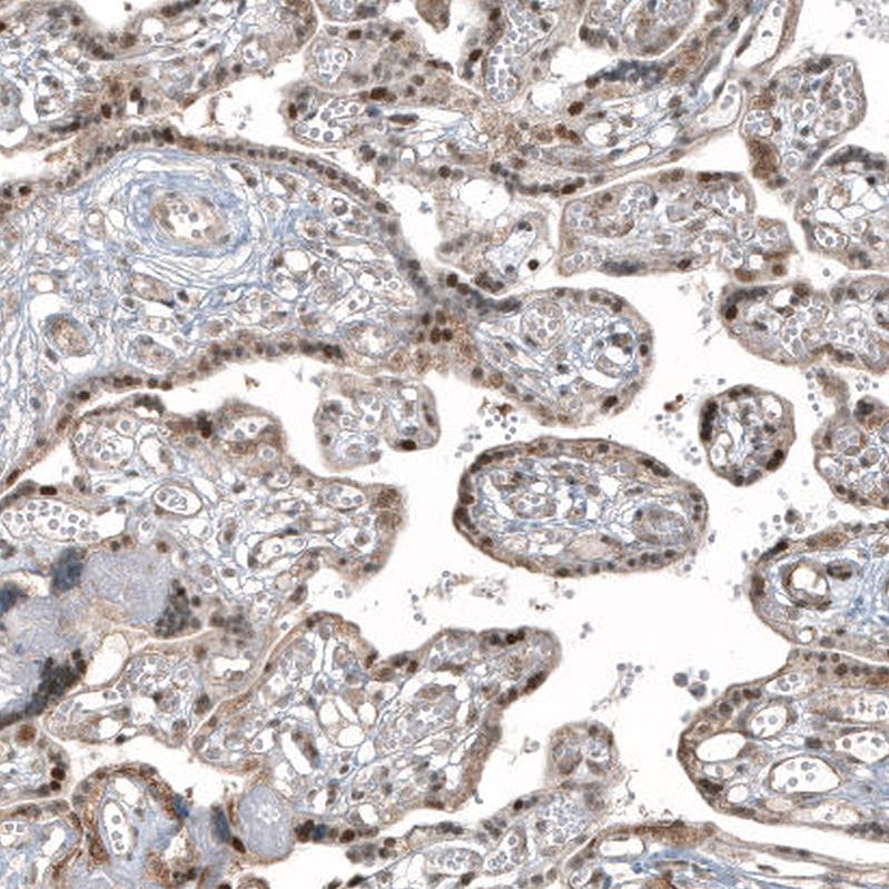
- Submitted by
- Atlas Antibodies (provider)
- Main image
- Experimental details
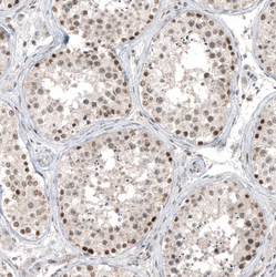
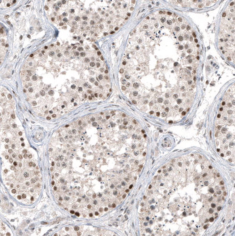
- Immunohistochemical staining of human testis shows moderate to strong nuclear positivity in cells in seminiferous ducts.
- Submitted by
- Atlas Antibodies (provider)
- Main image
- Experimental details
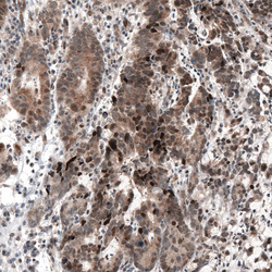
- Immunohistochemical staining of human stomach cancer shows moderate to strong cytoplasmic and nuclear positivity.

- Submitted by
- Atlas Antibodies (provider)
- Main image
- Experimental details
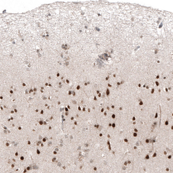
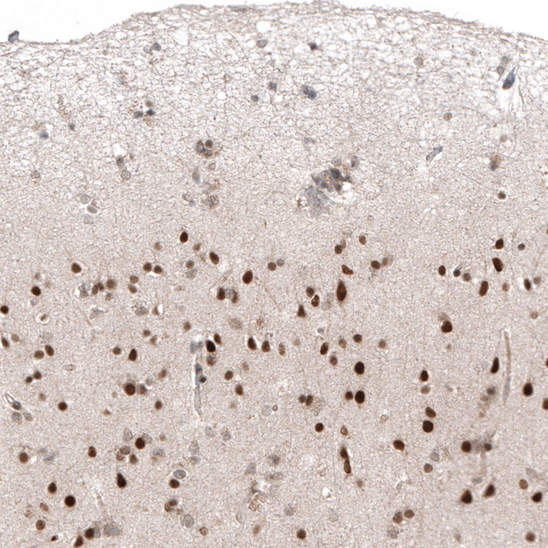
- Immunohistochemical staining of human cerebral cortex shows strong nuclear positivity in neurons.
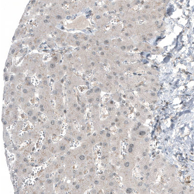
- Submitted by
- Atlas Antibodies (provider)
- Main image

- Experimental details
- Immunohistochemical staining of human liver shows no positivity in hepatocytes as expected.