Antibody data
- Antibody Data
- Antigen structure
- References [11]
- Comments [0]
- Validations
- Western blot [2]
- Other assay [10]
Submit
Validation data
Reference
Comment
Report error
- Product number
- 14-6008-93 - Provider product page

- Provider
- Invitrogen Antibodies
- Product name
- AIM2 Polyclonal Antibody, eBioscience™
- Antibody type
- Polyclonal
- Antigen
- Other
- Description
- Description: This polyclonal antibody reacts with human, mouse, and rat AIM2 (also known as Antigen isolated from Immunoselected Melanoma 2). A member of the HIN-2000 family, this 40-kDa cytoplasmic protein is expressed in the spleen, small intestine, peripheral blood lymphocytes, and testis. Moreover, AIM2 is expressed in tumors of the neuroectodermis, breast, ovary, and colon. As a component of the inflammosome, AIM2 detects double-stranded viral and bacterial DNA present in the cytosol and initiates Caspase1 activation. Expression of AIM2 can be induced by interferons. Applications Reported: This polyclonal antibody has been reported for use in immunoblotting (WB). Applications Tested: This polyclonal antibody has been tested by immunoblotting of Jurkat cells and mouse macrophages treated for 12 hrs with IFN gamma and LPS. This can be used at less than or equal to a 1:1000 dilution. It is recommended that the antibody be carefully titrated for optimal performance in the assay of interest. Purity: Greater than 90%, as determined by SDS-PAGE. Aggregation: Less than 10%, as determined by HPLC. Filtration: 0.2 µm post-manufacturing filtered.
- Reactivity
- Human, Mouse, Rat
- Host
- Rabbit
- Isotype
- IgG
- Vial size
- 100 µL
- Storage
- 4° C
Submitted references BECN2 (beclin 2) Negatively Regulates Inflammasome Sensors Through ATG9A-Dependent but ATG16L1- and LC3-Independent Non-Canonical Autophagy.
Inhibition of AIM2 inflammasome activation alleviates GSDMD-induced pyroptosis in early brain injury after subarachnoid haemorrhage.
MARK4 (Microtubule Affinity-Regulating Kinase 4)-Dependent Inflammasome Activation Promotes Atherosclerosis-Brief Report.
Epstein-Barr virus infection-induced inflammasome activation in human monocytes.
The PYHIN Protein p205 Regulates the Inflammasome by Controlling Asc Expression.
Acidification changes affect the inflammasome in human nucleus pulposus cells.
Expression analysis of inflammasomes in experimental models of inflammatory and fibrotic liver disease.
The AIM2 inflammasome is essential for host defense against cytosolic bacteria and DNA viruses.
The AIM2 inflammasome is critical for innate immunity to Francisella tularensis.
AIM2 recognizes cytosolic dsDNA and forms a caspase-1-activating inflammasome with ASC.
AIM2 activates the inflammasome and cell death in response to cytoplasmic DNA.
Deng G, Li C, Chen L, Xing C, Fu C, Qian C, Liu X, Wang HY, Zhu M, Wang RF
Autophagy 2022 Feb;18(2):340-356
Autophagy 2022 Feb;18(2):340-356
Inhibition of AIM2 inflammasome activation alleviates GSDMD-induced pyroptosis in early brain injury after subarachnoid haemorrhage.
Yuan B, Zhou XM, You ZQ, Xu WD, Fan JM, Chen SJ, Han YL, Wu Q, Zhang X
Cell death & disease 2020 Jan 30;11(1):76
Cell death & disease 2020 Jan 30;11(1):76
MARK4 (Microtubule Affinity-Regulating Kinase 4)-Dependent Inflammasome Activation Promotes Atherosclerosis-Brief Report.
Clement M, Chen X, Chenoweth HL, Teng Z, Thome S, Newland SA, Harrison J, Yu X, Finigan AJ, Mallat Z, Li X
Arteriosclerosis, thrombosis, and vascular biology 2019 Aug;39(8):1645-1651
Arteriosclerosis, thrombosis, and vascular biology 2019 Aug;39(8):1645-1651
Epstein-Barr virus infection-induced inflammasome activation in human monocytes.
Torii Y, Kawada JI, Murata T, Yoshiyama H, Kimura H, Ito Y
PloS one 2017;12(4):e0175053
PloS one 2017;12(4):e0175053
The PYHIN Protein p205 Regulates the Inflammasome by Controlling Asc Expression.
Ghosh S, Wallerath C, Covarrubias S, Hornung V, Carpenter S, Fitzgerald KA
Journal of immunology (Baltimore, Md. : 1950) 2017 Nov 1;199(9):3249-3260
Journal of immunology (Baltimore, Md. : 1950) 2017 Nov 1;199(9):3249-3260
Acidification changes affect the inflammasome in human nucleus pulposus cells.
Brand FJ 3rd, Forouzandeh M, Kaur H, Travascio F, de Rivero Vaccari JP
Journal of inflammation (London, England) 2016;13(1):29
Journal of inflammation (London, England) 2016;13(1):29
Expression analysis of inflammasomes in experimental models of inflammatory and fibrotic liver disease.
Boaru SG, Borkham-Kamphorst E, Tihaa L, Haas U, Weiskirchen R
Journal of inflammation (London, England) 2012 Nov 28;9(1):49
Journal of inflammation (London, England) 2012 Nov 28;9(1):49
The AIM2 inflammasome is essential for host defense against cytosolic bacteria and DNA viruses.
Rathinam VA, Jiang Z, Waggoner SN, Sharma S, Cole LE, Waggoner L, Vanaja SK, Monks BG, Ganesan S, Latz E, Hornung V, Vogel SN, Szomolanyi-Tsuda E, Fitzgerald KA
Nature immunology 2010 May;11(5):395-402
Nature immunology 2010 May;11(5):395-402
The AIM2 inflammasome is critical for innate immunity to Francisella tularensis.
Fernandes-Alnemri T, Yu JW, Juliana C, Solorzano L, Kang S, Wu J, Datta P, McCormick M, Huang L, McDermott E, Eisenlohr L, Landel CP, Alnemri ES
Nature immunology 2010 May;11(5):385-93
Nature immunology 2010 May;11(5):385-93
AIM2 recognizes cytosolic dsDNA and forms a caspase-1-activating inflammasome with ASC.
Hornung V, Ablasser A, Charrel-Dennis M, Bauernfeind F, Horvath G, Caffrey DR, Latz E, Fitzgerald KA
Nature 2009 Mar 26;458(7237):514-8
Nature 2009 Mar 26;458(7237):514-8
AIM2 activates the inflammasome and cell death in response to cytoplasmic DNA.
Fernandes-Alnemri T, Yu JW, Datta P, Wu J, Alnemri ES
Nature 2009 Mar 26;458(7237):509-13
Nature 2009 Mar 26;458(7237):509-13
No comments: Submit comment
Supportive validation
- Submitted by
- Invitrogen Antibodies (provider)
- Main image

- Experimental details
- Cell lysates prepared from Jurkat cells (lane 1) and mouse macrophages left untreated (lane 2) or treated for 12 hr with IFN gamma and LPS (lane 3) were immunoblotted with a 1:1000 dilution of the Anti-AIM2 polyclonal antibody. Bands were visualized using Anti-Rabbit IgG HRP.
- Submitted by
- Invitrogen Antibodies (provider)
- Main image

- Experimental details
- Cell lysates prepared from Jurkat cells (lane 1) and mouse macrophages left untreated (lane 2) or treated for 12 hr with IFN gamma and LPS (lane 3) were immunoblotted with a 1:1000 dilution of the Anti-AIM2 polyclonal antibody. Bands were visualized using Anti-Rabbit IgG HRP.
Supportive validation
- Submitted by
- Invitrogen Antibodies (provider)
- Main image

- Experimental details
- NULL
- Submitted by
- Invitrogen Antibodies (provider)
- Main image

- Experimental details
- NULL
- Submitted by
- Invitrogen Antibodies (provider)
- Main image

- Experimental details
- Figure 6 Hepatic expression of AIM2 and NALP4/NLRC4 in rats after administration of CCl 4 or BDL surgery. Liver specimen from animals that received CCl 4 or underwent BDL for indicated time points were stained with antibodies specific for AIM2 ( A ) or NALP4/NLRC4 ( B ). Section from control animals and a stain with an unspecific rabbit IgG served as controls in this analysis.
- Submitted by
- Invitrogen Antibodies (provider)
- Main image

- Experimental details
- Fig 2 Inflammasome-related protein and gene expression of THP-1 cells after EBV infection. (A) IL-1beta concentration in the supernatant of THP-1 cells, Jurkat cells, or BJAB cells at 48 hours after incubation with RPMI (mock), AGS-EBV-GFP cell supernatant (AGSGFP: 6.0x10 6 , 6.0x10 5 and 6.0x10 4 GBU/mL), or B95-8 cell supernatant (B95-8). The data represent two experiments each run with duplicate samples. The error bars represent S.E. The asterisk (*) indicates p < 0.001 based on comparison to the 0 h time-point by one-way ANOVA with Tukey's post hoc test. (B) IL-1beta concentration in the supernatant of THP-1 cells, pretreated with or without caspase-1 inhibitor, at 24 hours after incubation with RPMI, AGSGFP (6.0x10 6 GBU/mL) or LPS (10 ng/mL). The data represent two experiments each run with duplicate samples. The error bars represent S.E. The single asterisk (*) indicates p = 0.003 and the double asterisk (**) indicates p < 0.001 by one-way ANOVA with Tukey's post hoc test. (C) IL-1beta concentration in the supernatant of THP-1 cells over time, following incubation with 6.0x10 6 GBU/mL of AGSGFP supernatant or virion-free supernatant (filtration). The data represent one experiment with triplicate samples. The error bars represent S.E. The single asterisk (*) indicates p = 0.041 and the double asterisk (**) indicates p = 0.003 based on comparison to the 0 h time-point by one-way ANOVA with Tukey's post hoc test. (D) Immunoblot analysis of caspase-1, NLRP3,
- Submitted by
- Invitrogen Antibodies (provider)
- Main image

- Experimental details
- Fig 4 Inflammasome activation in AIM2 knockdown THP-1 cells after EBV infection. The following analyses were performed at 24 hours after incubation with RPMI (mock) or AGS-EBV-GFP cell supernatant (AGSGFP). (A) AIM2 mRNA expression of siRNA-treated THP-1 cells. The asterisk (*) indicates p = 0.02 (B) Immunoblot analysis of siRNA-treated THP-1 cells. beta-actin was blotted as a loading control. (C) mRNA expression of caspase-1 and IL-1beta in siRNA-treated THP-1 cells. The double asterisk (**) indicates p
- Submitted by
- Invitrogen Antibodies (provider)
- Main image

- Experimental details
- Fig 3 EBV infection and inflammasome activation in human monocytes. (A) Confocal microscopic images of human monocytes 48 h after incubation with RPMI medium (no infection) or an AGS-EBV-GFP cell supernatant (AGSGFP: 6.0x10 6 or 6.0x10 5 GBU/mL). (B) EBV gene expression of human monocytes at 48 h after incubation with 6.0x10 6 GBU/mL of an AGSGFP cell supernatant. EBV gene expression was quantified relative to beta-2-microglobulin expression. The data represent one experiment with triplicate samples. The error bars represent S.E. (C) IL-1beta concentration in the supernatant of human monocytes over time, following incubation with 6.0x10 6 GBU/mL of an AGSGFP cell supernatant. The data represent one experiment with duplicate samples. The error bars represent S.E. (D) Immunoblot analysis of caspase-1, IFI16, and AIM2 protein expression in human primary monocyte lysates over time, following incubation with 6.0x10 6 GBU/mL of an AGSGFP cell supernatant. (E) Inflammasome-related gene (IL-1beta, caspase-1, AIM2, NLRP3 and IFI16) expression of human monocytes over time, following incubation with 6.0x10 6 GBU/mL of an AGSGFP cell supernatant, was measured using RT-PCR. The data are expressed as fold changes compared to the 0-hour time-point. The data represent one experiment with triplicate samples. The error bars represent S.E. The asterisk (*) indicates p < 0.001 based on comparison to the 0-hour time-point by one-way ANOVA with Tukey's post hoc test.
- Submitted by
- Invitrogen Antibodies (provider)
- Main image

- Experimental details
- Fig. 2 The neuronal AIM2 inflammasome-mediated pyroptosis pathway was explored in the mouse temporal cortex after SAH. a Representative Nissl staining images of temporal cortex in sham, post-SAH 24 h and post-SAH 72 h groups; b Representative immunohistochemistry images of AIM2, GSDMD, caspase-1 and ASC in sham, post-SAH 24 h and post-SAH 72 h groups; c Western blot assay for the expression of AIM2, GSDMD, GSDMD-N, caspase-1, caspase-1 p20 and ASC in temporal cortex after SAH; d Quantification of AIM2, GSDMD, GSDMD-N, caspase-1, caspase-1 p20 and ASC. n = 6 per group. Bars represent the mean +- S.E.M. * P < 0.05, ** P < 0.01, *** P < 0.001, and ns means non-significant. Scale bars = 50 mum. GSDMD gasdermin D; GSDMD-N N-terminus of gasdermin D; ASC apoptosis-associated speck-like protein containing a CARD.
- Submitted by
- Invitrogen Antibodies (provider)
- Main image

- Experimental details
- Fig. 3 The AIM2 inflammasome-mediated pyroptosis pathway was measured in primary cortical neurons exposed to oxyHb. a Representative images of cultured primary cortical neurons, scale bars (NeuN) = 50 mum, scale bars (MAP2) = 200 mum; b Western blot assay for the expression of AIM2, GSDMD, GSDMD-N, caspase-1, caspase-1 p20 and ASC in all groups; c Quantification of AIM2, GSDMD, GSDMD-N, caspase-1, caspase-1 p20 and ASC; d , e Quantitative analysis of IL-1beta and IL-18 secreted in the supernatant; f Flow cytometry analysis of neurons in all groups; g Quantification of caspase-1 and PI-positive neurons in all groups. Bars represent the mean +- S.E.M. * P < 0.05, *** P < 0.001, **** P < 0.0001 and ns means non-significant. OxyHb oxyhaemoglobin; NeuN neuronal nuclei; MAP2 microtubule associated protein 2; IL-1beta interleukin-1beta; IL-18 interleukin-18; PI propidium iodide.
- Submitted by
- Invitrogen Antibodies (provider)
- Main image

- Experimental details
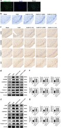
- Fig. 5 Effects of inhibited AIM2 inflammasome activation on GSDMD-induced pyroptosis 24 h post-SAH in vivo. a The transfection of LV-NCGFP in vivo, scale bars = 200 mum; b Representative immunohistochemistry images of AIM2, GSDMD, caspase-1 and ASC in all groups, scale bars = 50 mum; c Representative Nissl staining images of temporal cortex in all groups, scale bars = 50 mum; d , f Western blot assay for the expression of AIM2, GSDMD, GSDMD-N, caspase-1, caspase-1 p20 and ASC in all groups; e , g Quantification of AIM2, GSDMD, GSDMD-N, caspase-1, caspase-1 p20 and ASC. n = 6 per group. Bars represent the mean +- S.E.M. *** P < 0.001, and ns means non-significant. DAPI 4,6-diamidino-2-phenylindole.
- Submitted by
- Invitrogen Antibodies (provider)
- Main image

- Experimental details
- Fig. 6 Effects of inhibited AIM2 inflammasome activation on GSDMD-induced pyroptosis in primary cortical neurons exposed to oxyHb. a , c Western blot assay for the expression of AIM2, GSDMD, GSDMD-N, caspase-1, caspase-1 p20 and ASC in all groups; b , d Quantification of AIM2, GSDMD, GSDMD-N, caspase-1, caspase-1 p20 and ASC; e , f Quantitative analysis of IL-1beta and IL-18 secreted in the supernatant; g Flow cytometry analysis of neurons in all groups; h Quantification of Caspase-1 and PI-positive neurons in all groups. Bars represent the mean +- S.E.M. *** P < 0.001, **** P < 0.0001 and ns means non-significant.
 Explore
 Validate
 Learn
Learn Western blot