 Explore
 Validate
 Learn
LearnPA5-27076
antibody from Invitrogen Antibodies
Targeting: GPBAR1
BG37, GPCR, GPCR19, GPR131, M-BAR, MGC40597, TGR5
 Western blot
Antibody data
- Antibody Data
- Antigen structure
- References [1]
- Comments [0]
- Validations
- Western blot [1]
- Immunohistochemistry [3]
Submit
Validation data
Reference
Comment
Report error
- Product number
- PA5-27076 - Provider product page

- Provider
- Invitrogen Antibodies
- Product name
- TGR5 Polyclonal Antibody
- Antibody type
- Polyclonal
- Antigen
- Synthetic peptide
- Description
- Recommended positive controls: A431, H1299, HeLa, HepG2, Molt-4, Raji. Predicted reactivity: Chimpanzee (100%). Store product as a concentrated solution. Centrifuge briefly prior to opening the vial.
- Reactivity
- Human
- Host
- Rabbit
- Isotype
- IgG
- Vial size
- 100 µL
- Concentration
- 1 mg/mL
- Storage
- Store at 4°C short term. For long term storage, store at -20°C, avoiding freeze/thaw cycles.
Submitted references Enteroendocrine cells are a potential source of serum autotaxin in men.
Bolier R, Tolenaars D, Kremer AE, Saris J, Parés A, Verheij J, Bosma PJ, Beuers U, Oude Elferink RPJ
Biochimica et biophysica acta 2016 Apr;1862(4):696-704
Biochimica et biophysica acta 2016 Apr;1862(4):696-704
No comments: Submit comment
Supportive validation
- Submitted by
- Invitrogen Antibodies (provider)
- Main image

- Experimental details
- Western Blot using TGR5 Polyclonal Antibody (Product # PA5-27076). Sample (30 µg of whole cell lysate). Lane A: HepG2. 10% SDS PAGE. TGR5 Polyclonal Antibody (Product # PA5-27076) diluted at 1:1,000. The HRP-conjugated anti-rabbit IgG antibody was used to detect the primary antibody.

Supportive validation
- Submitted by
- Invitrogen Antibodies (provider)
- Main image
- Experimental details
- Immunohistochemistry analysis of TGR5 Antibody (Product # PA5-27076) was performed on a mouse (Mus musculus) adrenal gland. To expose target proteins, antigen retrieval was performed by microwaving tissues for 10 minutes in 10mM sodium citrate buffer (pH 6.0). Following antigen retrieval, endogenous peroxidases were blocked with 3% hydrogen peroxide for 10 min at room temperature. Tissue slides were washed with deionized water and TBST, and then blocked in a protein free blocking buffer for 7 min at room temperature. Tissues were incubated with a TGR5 Antibody (Product # PA5-27076) diluted 1:100 in distilled water or rabbit IgG as a negative control for 1 hour at room temperature in a humidified chamber. Tissues were washed extensively in TBST and detection was performed using a polymer, and then followed by colorimetric detection using a DAB kit. Tissues were counterstained with hematoxylin and dehydrated with ethanol and xylene to prep for mounting. Images were taken on a microscope at 20X magnification. In the image, immunolabeling is observed in the neurons of the adrenal gland medulla.

- Submitted by
- Invitrogen Antibodies (provider)
- Main image

- Experimental details
- Immunohistochemistry analysis of TGR5 Antibody (Product # PA5-27076) was performed on a mouse adrenal gland. Antigen retrieval was performed by microwaving tissues for 10 minutes in 10mM sodium citrate buffer (pH 6.0). Following antigen retrieval, endogenous peroxidases were blocked with 3% hydrogen peroxide for 10 min at room temperature. Tissue slides were washed with deionized water and TBST, and then blocked in a protein free blocking buffer for 7 min at room temperature. Tissues were incubated with a TGR5 Antibody (Product # PA5-27076) diluted 1:100 in distilled water or rabbit IgG as a negative control for 1 hour at room temperature in a humidified chamber. Tissues were washed extensively in TBST and detection was performed using a polymer, then followed by colorimetric detection using a DAB kit. Tissues were counterstained with hematoxylin and dehydrated with ethanol and xylene to prep for mounting. Images were taken at 20X magnification. In the image, immunolabeling is observed in the neurons of the adrenal gland medulla. Data courtesy of Antibody Data Exchange Program.

- Submitted by
- Invitrogen Antibodies (provider)
- Main image

- Experimental details
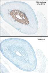
- Immunohistochemical analysis of paraffin-embedded HUH-7 xenograft, using TGR5 (Product # PA5-27076) antibody at 1:100 dilution. Antigen Retrieval: EDTA based buffer, pH 8.0, 15 min.
