Explore
 Validate
 Learn
Learn Western blot
Antibody data
- Antibody Data
- Antigen structure
- References [1]
- Comments [0]
- Validations
- Western blot [2]
- Immunocytochemistry [1]
- Immunohistochemistry [5]
Submit
Validation data
Reference
Comment
Report error
- Product number
- HPA028024 - Provider product page

- Provider
- Atlas Antibodies
- Proper citation
- Atlas Antibodies Cat#HPA028024, RRID:AB_10603787
- Product name
- Anti-COPA
- Antibody type
- Polyclonal
- Reactivity
- Human, Mouse, Rat
- Host
- Rabbit
- Conjugate
- Unconjugated
- Antigen sequence
VHGNMLHYVKDRFLRQLDFNSSKDVAVMQLRSGSK
FPVFNMSYNPAENAVLLCTRASNLENSTYDLYTIP
KDADSQNPDAPEGKRSSGLTAVWVARN- Isotype
- IgG
- Vial size
- 100 µl
- Storage
- Store at +4°C for short term storage. Long time storage is recommended at -20°C.
Submitted references COPA mutations impair ER-Golgi transport and cause hereditary autoimmune-mediated lung disease and arthritis
Watkin L, Jessen B, Wiszniewski W, Vece T, Jan M, Sha Y, Thamsen M, Santos-Cortez R, Lee K, Gambin T, Forbes L, Law C, Stray-Pedersen A, Cheng M, Mace E, Anderson M, Liu D, Tang L, Nicholas S, Nahmod K, Makedonas G, Canter D, Kwok P, Hicks J, Jones K, Penney S, Jhangiani S, Rosenblum M, Dell S, Waterfield M, Papa F, Muzny D, Zaitlen N, Leal S, Gonzaga-Jauregui C, Boerwinkle E, Eissa N, Gibbs R, Lupski J, Orange J, Shum A
Nature Genetics 2015 April;47(6):654-660
Nature Genetics 2015 April;47(6):654-660
No comments: Submit comment
Supportive validation
- Submitted by
- Atlas Antibodies (provider)
- Main image
- Experimental details
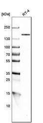
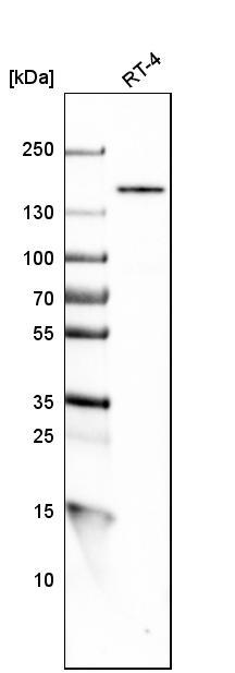
- Western blot analysis in human cell line RT-4.
- Submitted by
- Atlas Antibodies (provider)
- Main image
- Experimental details
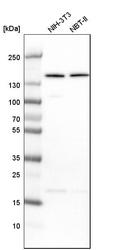
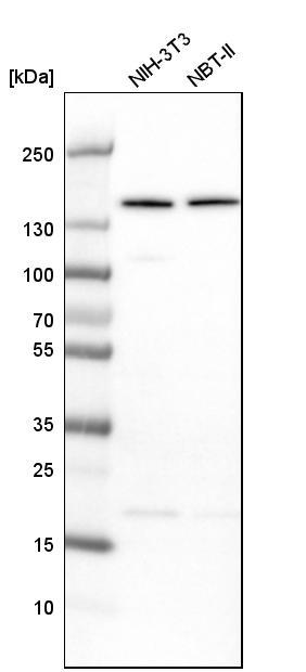
- Western blot analysis in mouse cell line NIH-3T3 and rat cell line NBT-II.
Supportive validation
- Submitted by
- Atlas Antibodies (provider)
- Main image
- Experimental details
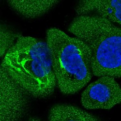
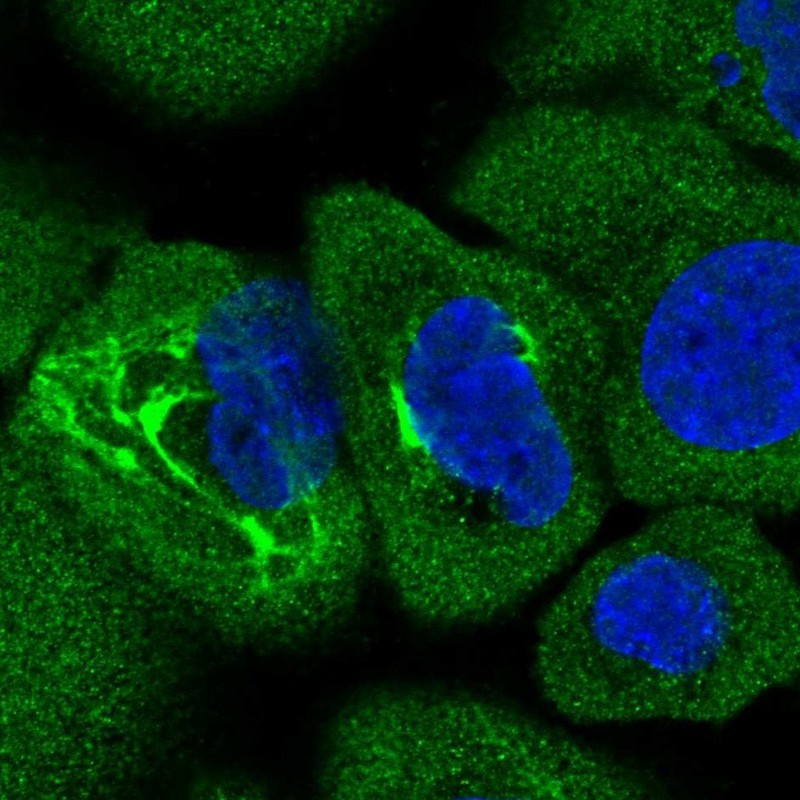
- Immunofluorescent staining of human cell line A-431 shows positivity in cytoplasm, intermediate filaments & nucleus but excluded from the nucleoli.
- Sample type
- HUMAN
Supportive validation
- Submitted by
- Atlas Antibodies (provider)
- Main image

- Experimental details
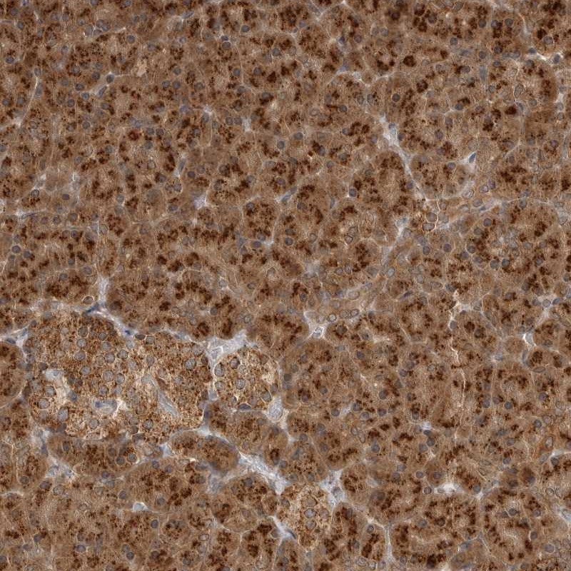
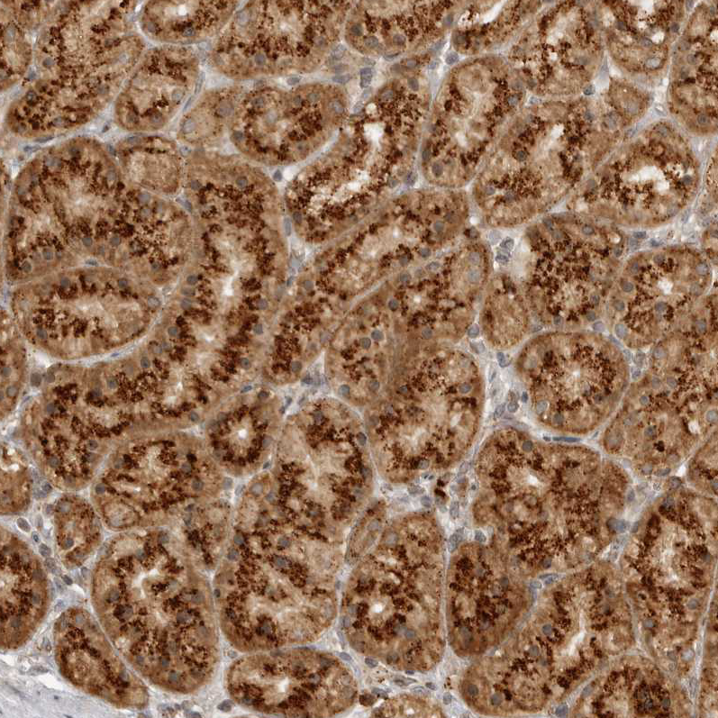
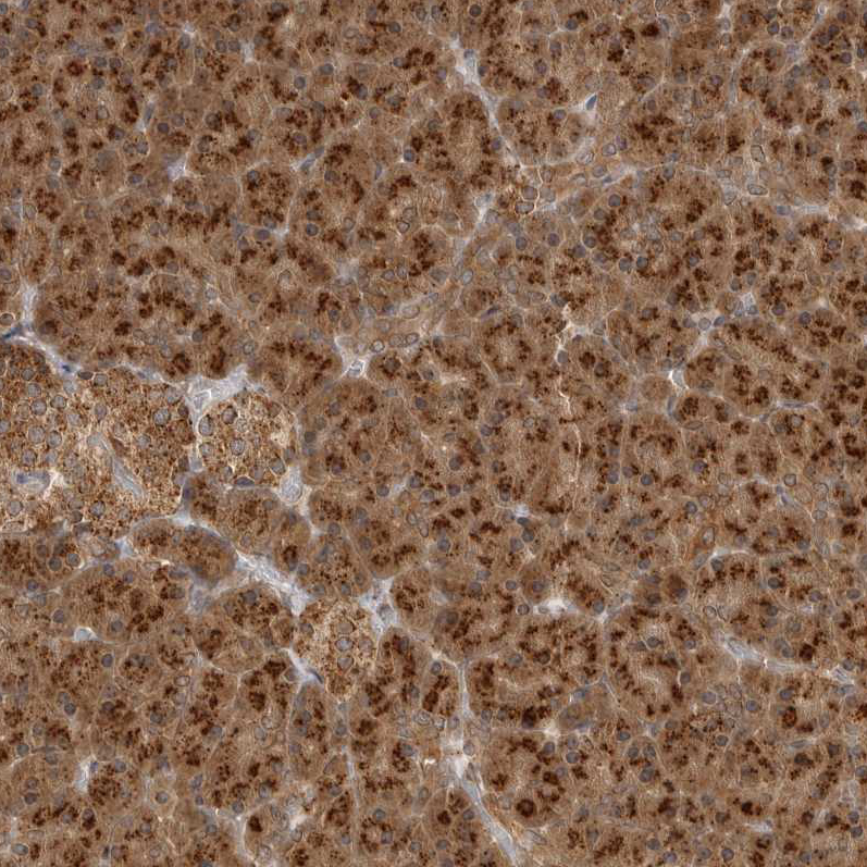
- Immunohistochemical staining of human pancreas shows strong cytoplasmic positivity with a granular pattern in exocrine cells.
- Submitted by
- Atlas Antibodies (provider)
- Main image

- Experimental details
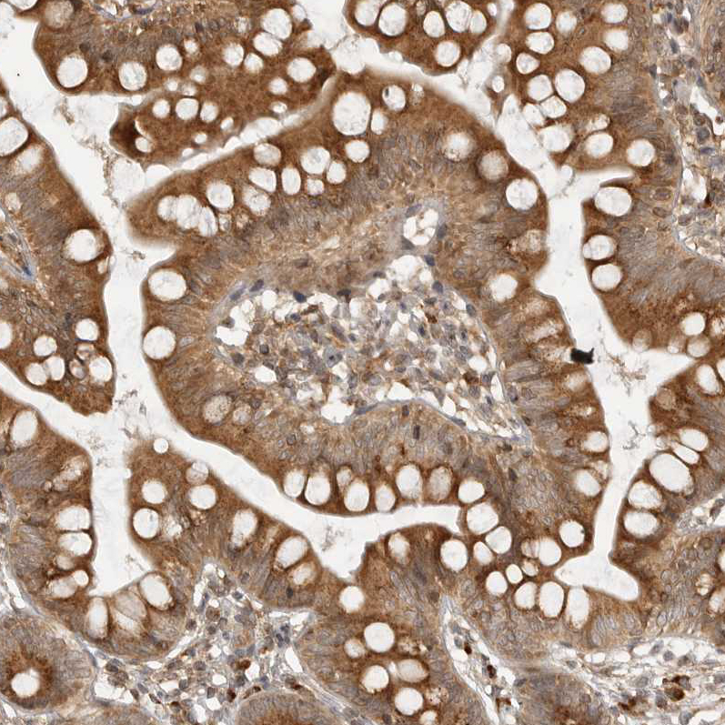
- Immunohistochemical staining of human small intestine shows moderate cytoplasmic positivity in glandular cells.
- Sample type
- HUMAN
- Submitted by
- Atlas Antibodies (provider)
- Main image
- Experimental details
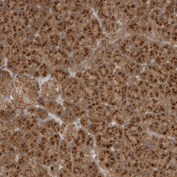
- Immunohistochemical staining of human stomach shows moderate granular cytoplasmic positivity in glandular cells.
- Sample type
- HUMAN
- Submitted by
- Atlas Antibodies (provider)
- Main image
- Experimental details
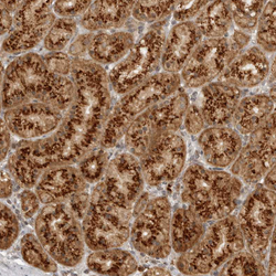
- Immunohistochemical staining of human pancreas shows moderate to strong granular cytoplasmic positivity in exocrine glandular cells.
- Sample type
- HUMAN
- Submitted by
- Atlas Antibodies (provider)
- Main image

- Experimental details
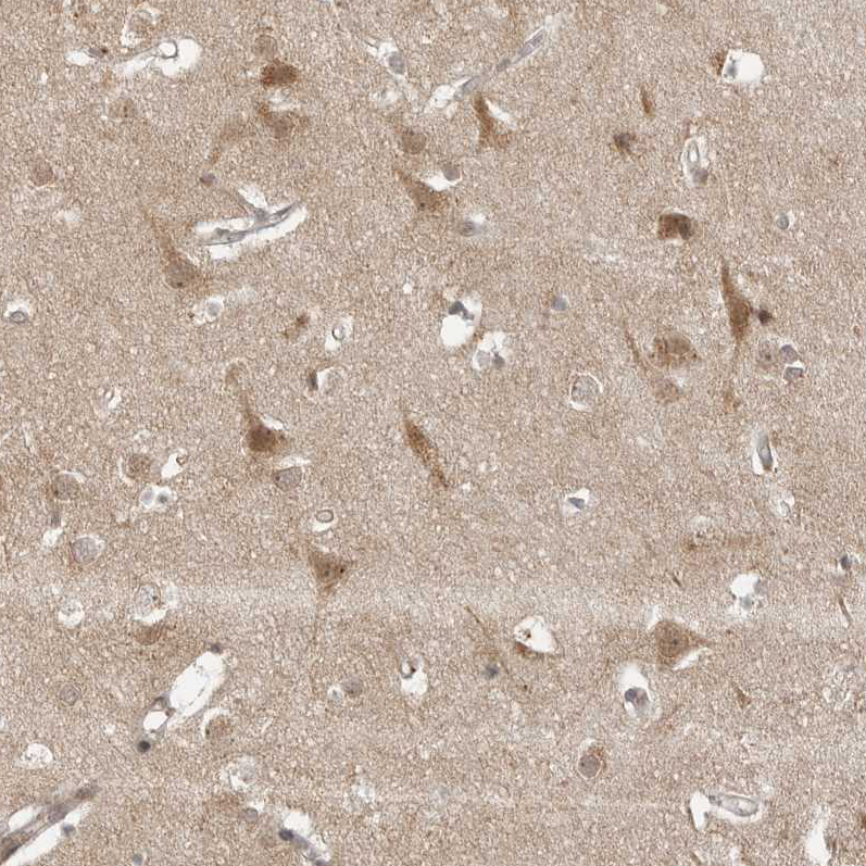
- Immunohistochemical staining of human cerebral cortex shows moderate cytoplasmic positivity in neurons.
- Sample type
- HUMAN