 Explore
 Validate
 Learn
LearnMA5-15142
antibody from Invitrogen Antibodies
Targeting: BCL2L1
Bcl-X, bcl-xL, bcl-xS, BCL2L, BCLX, PPP1R52
 Western blot
 Immunoprecipitation
 Flow cytometry
Antibody data
- Antibody Data
- Antigen structure
- References [6]
- Comments [0]
- Validations
- Western blot [3]
- Immunohistochemistry [1]
- Other assay [5]
Submit
Validation data
Reference
Comment
Report error
- Product number
- MA5-15142 - Provider product page

- Provider
- Invitrogen Antibodies
- Product name
- Bcl-xL Monoclonal Antibody (C.85.1)
- Antibody type
- Monoclonal
- Antigen
- Synthetic peptide
- Description
- It is not recommended to aliquot this antibody. This antibody is not cross-reactive with other Bcl-2 family members.
- Reactivity
- Human, Mouse, Rat
- Host
- Rabbit
- Isotype
- IgG
- Antibody clone number
- C.85.1
- Vial size
- 100 µL
- Concentration
- 144 µg/mL
- Storage
- -20°C
Submitted references Acetone Extract of Cornus officinalis Leaves Exerts Anti-Melanoma Effects via Inhibiting STAT3 Signaling.
MicroRNA-325-3p Facilitates Immune Escape of Mycobacterium tuberculosis through Targeting LNX1 via NEK6 Accumulation to Promote Anti-Apoptotic STAT3 Signaling.
Protective Effect of a Fucose-Rich Fucoidan Isolated from Saccharina japonica against Ultraviolet B-Induced Photodamage In Vitro in Human Keratinocytes and In Vivo in Zebrafish.
Activation of prolyl hydroxylase-2 for stabilization of mitochondrial stress along with simultaneous downregulation of HIF-1α/FASN in ER + breast cancer subtype.
ALA-mediated biphasic downregulation of α-7nAchR/HIF-1α along with mitochondrial stress modulation strategy in mammary gland chemoprevention.
Alpha-linolenic acid stabilizes HIF-1 α and downregulates FASN to promote mitochondrial apoptosis for mammary gland chemoprevention.
Xu R, Zeng M, Wu Y, Wang S, Zhang B, Zhang J, Kan Y, Li B, Cao B, Zheng X, Feng W
OncoTargets and therapy 2021;14:3487-3501
OncoTargets and therapy 2021;14:3487-3501
MicroRNA-325-3p Facilitates Immune Escape of Mycobacterium tuberculosis through Targeting LNX1 via NEK6 Accumulation to Promote Anti-Apoptotic STAT3 Signaling.
Fu B, Xue W, Zhang H, Zhang R, Feldman K, Zhao Q, Zhang S, Shi L, Pavani KC, Nian W, Lin X, Wu H
mBio 2020 Jun 2;11(3)
mBio 2020 Jun 2;11(3)
Protective Effect of a Fucose-Rich Fucoidan Isolated from Saccharina japonica against Ultraviolet B-Induced Photodamage In Vitro in Human Keratinocytes and In Vivo in Zebrafish.
Su W, Wang L, Fu X, Ni L, Duan D, Xu J, Gao X
Marine drugs 2020 Jun 15;18(6)
Marine drugs 2020 Jun 15;18(6)
Activation of prolyl hydroxylase-2 for stabilization of mitochondrial stress along with simultaneous downregulation of HIF-1α/FASN in ER + breast cancer subtype.
Devi U, Singh M, Roy S, Gupta PS, Ansari MN, Saeedan AS, Kaithwas G
Cell biochemistry and function 2019 Jun;37(4):216-227
Cell biochemistry and function 2019 Jun;37(4):216-227
ALA-mediated biphasic downregulation of α-7nAchR/HIF-1α along with mitochondrial stress modulation strategy in mammary gland chemoprevention.
Roy S, Singh M, Sammi SR, Pandey R, Kaithwas G
Journal of cellular physiology 2019 Apr;234(4):4015-4029
Journal of cellular physiology 2019 Apr;234(4):4015-4029
Alpha-linolenic acid stabilizes HIF-1 α and downregulates FASN to promote mitochondrial apoptosis for mammary gland chemoprevention.
Roy S, Rawat AK, Sammi SR, Devi U, Singh M, Gautam S, Yadav RK, Rawat JK, Singh L, Ansari MN, Saeedan AS, Pandey R, Kumar D, Kaithwas G
Oncotarget 2017 Sep 19;8(41):70049-70071
Oncotarget 2017 Sep 19;8(41):70049-70071
No comments: Submit comment
Supportive validation
- Submitted by
- Invitrogen Antibodies (provider)
- Main image

- Experimental details
- Knockdown of Bcl-xL was achieved by transfecting Hep G2 cells with Bcl-xL specific validated siRNAs (Silencer® select Product # s1920). Western blot analysis (Fig. a) was performed using whole cell extracts from the Bcl-xL knockdown cells (lane 3), non-specific scrambled siRNA transfected cells (lane 2) and untransfected cells (lane 1). The blots were probed with Bcl-xL Monoclonal Antibody (Product # MA5-15142, 1:1000 dilution) and Goat anti-Rabbit IgG (H+L) Superclonal™ Secondary Antibody, HRP conjugate (Product # A27036, 0.25 µg/mL, 1:4000 dilution). Densitometric analysis of this western blot is shown in histogram (Fig. b). Decrease in signal upon siRNA mediated knock down confirms that antibody is specific to Bcl-xL.

- Submitted by
- Invitrogen Antibodies (provider)
- Main image

- Experimental details
- Western blot analysis was performed on whole cell extracts (30 µg lysate) of HeLa (Lane 1), Hep G2 (Lane 2), HCT-116 (Lane 3), A431 (Lane 4), U-87 MG (Lane 5), PC-12 (Lane 6) and Tissue extracts of Mouse brain (Lane 7) and Rat brain (Lane 8). The blot was probed with anti-Bcl-xL Monoclonal Antibody (Product # MA5-15142, 1:1000 dilution) and detected by chemiluminescence using Goat anti-Rabbit IgG (H+L) Superclonal™ Secondary Antibody, HRP conjugate (Product # A27036, 0.25 µg/mL, 1:4000 dilution). A 26 kDa band corresponding to Bcl-xL was observed across all the cell lines and tissues tested.

- Submitted by
- Invitrogen Antibodies (provider)
- Main image

- Experimental details
- CRISPR-Cas9 mediated genome editing of BCL2L1 (as confirmed by next generation sequencing) was achieved by using LentiArray™ Lentiviral sgRNA (Product # A32042, AssayID CRISPR962685_LV) and LentiArray Cas9 Lentivirus (Product # A32064). Fig (a) Western blot analysis of BCL2L1 was performed by loading 30 µg of HeLa Wild Type (Lane 1), HeLa CAS9 (Lane 2) and HeLa CAS9 cells transduced with BCL2L1 Lentiviral sgRNA (Lane 3) whole cell extracts. The samples were electrophoresed using NuPAGE™ Novex™ 4-12% Bis-Tris Protein Gel (Product # NP0321BOX). Resolved proteins were then transferred onto a nitrocellulose membrane (Product # IB23001) by iBlot® 2 Dry Blotting System (Product # IB21001). The blot was probed with Anti-Bcl-xL Monoclonal Antibody (C.85.1) (Product # MA5-15142) using 1:1000 dilution and Goat anti-Rabbit IgG (H+L) Superclonal™ Recombinant Secondary Antibody, HRP (Product # A27036, 1:4000 dilution). Chemiluminescent detection was performed using SuperSignal™ West Dura Extended Duration Substrate (Product # 34076). A reduced signal in sgRNA transduced cells using the LentiArray™ CRISPR product line confirms that antibody is specific to BCL2L1 (Fig (b)).

Supportive validation
- Submitted by
- Invitrogen Antibodies (provider)
- Main image

- Experimental details
- Immunohistochemical analysis of Bcl-xL in paraffin-embedded human lung carcinoma using a Bcl-xL monoclonal antibody (Product # MA5-15142).

Supportive validation
- Submitted by
- Invitrogen Antibodies (provider)
- Main image
- Experimental details
- Figure 11 ALA mediated activation of mitochondrial associated protein signaling in mammary gland cells Protein extracted from individual groups [1-control, 2-DMBA treated, 3-ALA (0.25 ml/kg, p.o. + DMBA 8 mg/kg, i.v.) and 4- ALA (0.5 ml/kg, p.o. + DMBA 8 mg/kg, i.v.)] were subjected to immunoblotting of proapoptotic (BAX) and anti-apoptotic (Bcl-2 and Bcl-xl) protein with downstream apoptotic markers (VDAC, cytochrome-c, Apaf-1and procaspase9) of respective pathway. mRNA expression of above mentioned protein were also in line with the findings of immunoblotting assay. beta-actin was used as loading control. Each experiment was performed in triplicate. Values are presented as mean +- SD. Comparisons are made by the one-way ANOVA followed by Bonferroni multiple test. All groups are compared to the DMBA treated group (*p

- Submitted by
- Invitrogen Antibodies (provider)
- Main image

- Experimental details
- Immunoprecipitation of BcL-xL was performed on U-2 OS cells. Antigen-antibody complexes were formed by incubating 750 µg of U-2 OS whole cell lysate with 4 µg of a BcL-xL monoclonal antibody (Product # MA5-11950) or Mouse IgG2a isotype control (Product # MA1-10418) overnight on a rocking platform at 4°C. The immune complexes were captured on 100 µL Protein A/G Agarose (Product # 20421), washed extensively, and eluted with 5X Lane Marker Reducing Sample Buffer (Product # 39000). The IP samples and 25 µg of U-2 OS whole cell lysate (as a positive control for Western blot detection) were resolved on a 4-20% Tris-HCl polyacrylamide gel, transferred to a PVDF membrane, and blocked with 5% BSA/TBS-0.1%Tween for at least 1 hour. The membrane was probed with a BcL-xL rabbit monoclonal antibody (Product # MA5-15142) at a dilution of 1:1000 overnight rotating at 4°C, washed in TBST, and probed with an HRP-conjugated mouse anti-rabbit light chain secondary antibody at a dilution of 1:40,000 for at least 1 hour. Chemiluminescent detection was performed using SuperSignal West Pico (Product # 34080).

- Submitted by
- Invitrogen Antibodies (provider)
- Main image

- Experimental details
- Immunoprecipitation of Bcl-xL using a monoclonal antibody (Product # MA5-15142).

- Submitted by
- Invitrogen Antibodies (provider)
- Main image

- Experimental details
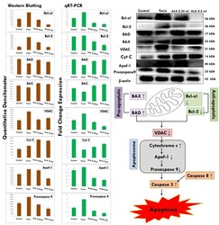
- 4 BBAP-2-mediated activation of mitochondrial associated protein signalling in mammary gland cells. Protein extracted from individual groups (group 1--control [0.9% normal saline, p.o. ]; group 2--toxic control [MNU, 8 mg/kg, i.v. ]; group 3--BBAP-2 + MNU [68.52 mug/kg, s.c. + 8 mg/kg MNU, i.v. ]; and group 4--BBAP-2 + MNU [137.04 mug/kg, s.c. + 8 mg/kg MNU, i.v.]) was subjected to immunoblotting of antiapoptotic (Bcl-2 and Bcl-xl) and proapoptotic (BAX) proteins with downstream apoptotic markers (VDAC, cytochrome-c, Apaf-1, and procaspase 9) of mitochondrial mediated apoptotic pathway. mRNA expression of above-mentioned protein was also done by real-time quantitative reverse transcription polymerase chain reaction (qRT-PCR). beta-actin was used as internal loading control. Treatment with BBAP-2 decreased the expression of antiapoptotic protein Bcl-xl without imparting any significant effect upon Bcl-2 and BAX (proapoptotic) while induction (BAX) and inhibition (Bcl-xl and Bcl-2) were observed when quantified through qRT-PCR assay. Each experiment was performed in triplicate. Values are presented as mean +- SD. Each group contains eight animals. Comparisons are made on the basis of the one-way ANOVA followed by Bonferroni multiple test. All groups are compared with the toxic control group (* p< 0.05, ** p< 0.01, *** p< 0.001)

- Submitted by
- Invitrogen Antibodies (provider)
- Main image

- Experimental details
- 3 Effect of ALA upon mitochondrial mediated death apoptosis pathway. Protein was extracted from individual groups (1, control; 2, MNU treated; and 3, ALA [0.25 ml/kg, p.o. + MNU47 mg/kg, i.v.]) and subjected to immunoblotting of proapoptotic (BAD) and antiapoptotic (Bcl-2 and Bcl-xl) proteins with its downstream apoptotic markers (VDAC, cytochrome-c, Apaf-1, and pro-caspase-9). mRNA expression of above mentioned proteins were also in line with the findings of immunoblotting assay. beta-Actin was used as loading control. Each experiment was performed three times. Values are presented as mean +- SD . Comparisons are made by one-way analysis of variance followed by Bonferroni's multiple test. All groups are compared with the MNU-treated group (* p < 0.05, ** p < 0.01, and *** p < 0.001). ALA: alpha linolenic acid; mRNA: messenger RNA; MNU: N -methyl- N -nitrosourea; VDAC: voltage-dependent anion channel [Color figure can be viewed at wileyonlinelibrary.com]
