Explore
 Validate
 Learn
Learn Western blot
 Immunocytochemistry
Antibody data
- Antibody Data
- Antigen structure
- References [21]
- Comments [0]
- Validations
- Western blot [8]
- Immunocytochemistry [3]
- Immunoprecipitation [2]
- Immunohistochemistry [13]
- Flow cytometry [1]
- Chromatin Immunoprecipitation [1]
Submit
Validation data
Reference
Comment
Report error
- Product number
- GTX101435 - Provider product page

- Provider
- GeneTex
- Proper citation
- GeneTex Cat#GTX101435, RRID:AB_1950062
- Product name
- beta Catenin antibody [N1N2-2], N-term
- Antibody type
- Polyclonal
- Reactivity
- Human, Mouse, Rat
- Host
- Rabbit
Submitted references Small G protein signalling modulator 2 (SGSM2) is involved in oestrogen receptor-positive breast cancer metastasis through enhancement of migratory cell adhesion via interaction with E-cadherin.
Suppression of SEMA6C promotes preantral follicles atresia with decreased cell junctions in mice ovaries.
Deficiency in STAT1 Signaling Predisposes Gut Inflammation and Prompts Colorectal Cancer Development.
Mechanisms of Matrix-Induced Chemoresistance of Breast Cancer Cells-Deciphering Novel Potential Targets for a Cell Sensitization.
Targeting the Wnt/β-catenin pathway in human osteosarcoma cells.
LncTIC1 interacts with β-catenin to drive liver TIC self-renewal and liver tumorigenesis.
Integrin Activation Contributes to Lower Cisplatin Sensitivity in MV3 Melanoma Cells by Inducing the Wnt Signalling Pathway.
Cytoplasmic aryl hydrocarbon receptor regulates glycogen synthase kinase 3 beta, accelerates vimentin degradation, and suppresses epithelial-mesenchymal transition in non-small cell lung cancer cells.
The phosphorylation status of T522 modulates tissue-specific functions of SIRT1 in energy metabolism in mice.
Control of the negative IRES trans-acting factor KHSRP by ubiquitination.
Antroquinonol, a Ubiquinone Derivative from the Mushroom Antrodia camphorata, Inhibits Colon Cancer Stem Cell-like Properties: Insights into the Molecular Mechanism and Inhibitory Targets.
Antroquinonol Targets FAK-Signaling Pathway Suppressed Cell Migration, Invasion, and Tumor Growth of C6 Glioma.
Inhibition of colon cancer cell growth by nanoemulsion carrying gold nanoparticles and lycopene.
WNT10B enhances proliferation through β-catenin and RAC1 GTPase in human corneal endothelial cells.
Involvement of RARRES3 in the regulation of Wnt proteins acylation and signaling activities in human breast cancer cells.
SH3BGRL3 Protein as a Potential Prognostic Biomarker for Urothelial Carcinoma: A Novel Binding Partner of Epidermal Growth Factor Receptor.
Pigment epithelium-derived factor 34-mer peptide prevents liver fibrosis and hepatic stellate cell activation through down-regulation of the PDGF receptor.
Interleukin-1β-induced Wnt5a enhances human corneal endothelial cell migration through regulation of Cdc42 and RhoA.
SERPINB3 is associated with TGF-β1 and cytoplasmic β-catenin expression in hepatocellular carcinomas with poor prognosis.
Loss of liver E-cadherin induces sclerosing cholangitis and promotes carcinogenesis.
Antimigratory Effects of the Methanol Extract from Momordica charantia on Human Lung Adenocarcinoma CL1 Cells.
Lin JH, Lee WJ, Wu HC, Wu CH, Chen LC, Huang CC, Chang HL, Cheng TC, Chang HW, Ho CT, Tu SH, Ho YS
Cell adhesion & migration 2019 Dec;13(1):120-137
Cell adhesion & migration 2019 Dec;13(1):120-137
Suppression of SEMA6C promotes preantral follicles atresia with decreased cell junctions in mice ovaries.
Yan W, Zhou S, Shen W, Cheng J, Yuan S, Ye S, Jin Y, Luo A, Wang S
Journal of cellular physiology 2019 Apr;234(4):4934-4943
Journal of cellular physiology 2019 Apr;234(4):4934-4943
Deficiency in STAT1 Signaling Predisposes Gut Inflammation and Prompts Colorectal Cancer Development.
Leon-Cabrera S, Vázquez-Sandoval A, Molina-Guzman E, Delgado-Ramirez Y, Delgado-Buenrostro NL, Callejas BE, Chirino YI, Pérez-Plasencia C, Rodríguez-Sosa M, Olguín JE, Salinas C, Satoskar AR, Terrazas LI
Cancers 2018 Sep 19;10(9)
Cancers 2018 Sep 19;10(9)
Mechanisms of Matrix-Induced Chemoresistance of Breast Cancer Cells-Deciphering Novel Potential Targets for a Cell Sensitization.
Jakubzig B, Baltes F, Henze S, Schlesinger M, Bendas G
Cancers 2018 Dec 6;10(12)
Cancers 2018 Dec 6;10(12)
Targeting the Wnt/β-catenin pathway in human osteosarcoma cells.
Fang F, VanCleave A, Helmuth R, Torres H, Rickel K, Wollenzien H, Sun H, Zeng E, Zhao J, Tao J
Oncotarget 2018 Dec 4;9(95):36780-36792
Oncotarget 2018 Dec 4;9(95):36780-36792
LncTIC1 interacts with β-catenin to drive liver TIC self-renewal and liver tumorigenesis.
Chen Z, Yao L, Liu Y, Zhu P
Cancer letters 2018 Aug 28;430:88-96
Cancer letters 2018 Aug 28;430:88-96
Integrin Activation Contributes to Lower Cisplatin Sensitivity in MV3 Melanoma Cells by Inducing the Wnt Signalling Pathway.
Piva MBR, Jakubzig B, Bendas G
Cancers 2017 Sep 16;9(9)
Cancers 2017 Sep 16;9(9)
Cytoplasmic aryl hydrocarbon receptor regulates glycogen synthase kinase 3 beta, accelerates vimentin degradation, and suppresses epithelial-mesenchymal transition in non-small cell lung cancer cells.
Li CH, Liu CW, Tsai CH, Peng YJ, Yang YH, Liao PL, Lee CC, Cheng YW, Kang JJ
Archives of toxicology 2017 May;91(5):2165-2178
Archives of toxicology 2017 May;91(5):2165-2178
The phosphorylation status of T522 modulates tissue-specific functions of SIRT1 in energy metabolism in mice.
Lu J, Xu Q, Ji M, Guo X, Xu X, Fargo DC, Li X
EMBO reports 2017 May;18(5):841-857
EMBO reports 2017 May;18(5):841-857
Control of the negative IRES trans-acting factor KHSRP by ubiquitination.
Kung YA, Hung CT, Chien KY, Shih SR
Nucleic acids research 2017 Jan 9;45(1):271-287
Nucleic acids research 2017 Jan 9;45(1):271-287
Antroquinonol, a Ubiquinone Derivative from the Mushroom Antrodia camphorata, Inhibits Colon Cancer Stem Cell-like Properties: Insights into the Molecular Mechanism and Inhibitory Targets.
Lin HC, Lin MH, Liao JH, Wu TH, Lee TH, Mi FL, Wu CH, Chen KC, Cheng CH, Lin CW
Journal of agricultural and food chemistry 2017 Jan 11;65(1):51-59
Journal of agricultural and food chemistry 2017 Jan 11;65(1):51-59
Antroquinonol Targets FAK-Signaling Pathway Suppressed Cell Migration, Invasion, and Tumor Growth of C6 Glioma.
Thiyagarajan V, Tsai MJ, Weng CF
PloS one 2015;10(10):e0141285
PloS one 2015;10(10):e0141285
Inhibition of colon cancer cell growth by nanoemulsion carrying gold nanoparticles and lycopene.
Huang RF, Wei YJ, Inbaraj BS, Chen BH
International journal of nanomedicine 2015;10:2823-46
International journal of nanomedicine 2015;10:2823-46
WNT10B enhances proliferation through β-catenin and RAC1 GTPase in human corneal endothelial cells.
Lee JG, Heur M
The Journal of biological chemistry 2015 Oct 30;290(44):26752-64
The Journal of biological chemistry 2015 Oct 30;290(44):26752-64
Involvement of RARRES3 in the regulation of Wnt proteins acylation and signaling activities in human breast cancer cells.
Hsu TH, Jiang SY, Chang WL, Eckert RL, Scharadin TM, Chang TC
Cell death and differentiation 2015 May;22(5):801-14
Cell death and differentiation 2015 May;22(5):801-14
SH3BGRL3 Protein as a Potential Prognostic Biomarker for Urothelial Carcinoma: A Novel Binding Partner of Epidermal Growth Factor Receptor.
Chiang CY, Pan CC, Chang HY, Lai MD, Tzai TS, Tsai YS, Ling P, Liu HS, Lee BF, Cheng HL, Ho CL, Chen SH, Chow NH
Clinical cancer research : an official journal of the American Association for Cancer Research 2015 Dec 15;21(24):5601-11
Clinical cancer research : an official journal of the American Association for Cancer Research 2015 Dec 15;21(24):5601-11
Pigment epithelium-derived factor 34-mer peptide prevents liver fibrosis and hepatic stellate cell activation through down-regulation of the PDGF receptor.
Tsai TH, Shih SC, Ho TC, Ma HI, Liu MY, Chen SL, Tsao YP
PloS one 2014;9(4):e95443
PloS one 2014;9(4):e95443
Interleukin-1β-induced Wnt5a enhances human corneal endothelial cell migration through regulation of Cdc42 and RhoA.
Lee JG, Heur M
Molecular and cellular biology 2014 Sep 15;34(18):3535-45
Molecular and cellular biology 2014 Sep 15;34(18):3535-45
SERPINB3 is associated with TGF-β1 and cytoplasmic β-catenin expression in hepatocellular carcinomas with poor prognosis.
Turato C, Vitale A, Fasolato S, Ruvoletto M, Terrin L, Quarta S, Ramirez Morales R, Biasiolo A, Zanus G, Zali N, Tan PS, Hoshida Y, Gatta A, Cillo U, Pontisso P
British journal of cancer 2014 May 27;110(11):2708-15
British journal of cancer 2014 May 27;110(11):2708-15
Loss of liver E-cadherin induces sclerosing cholangitis and promotes carcinogenesis.
Nakagawa H, Hikiba Y, Hirata Y, Font-Burgada J, Sakamoto K, Hayakawa Y, Taniguchi K, Umemura A, Kinoshita H, Sakitani K, Nishikawa Y, Hirano K, Ikenoue T, Ijichi H, Dhar D, Shibata W, Akanuma M, Koike K, Karin M, Maeda S
Proceedings of the National Academy of Sciences of the United States of America 2014 Jan 21;111(3):1090-5
Proceedings of the National Academy of Sciences of the United States of America 2014 Jan 21;111(3):1090-5
Antimigratory Effects of the Methanol Extract from Momordica charantia on Human Lung Adenocarcinoma CL1 Cells.
Hsu HY, Lin JH, Li CJ, Tsang SF, Tsai CH, Chyuan JH, Chiu SJ, Chuang SE
Evidence-based complementary and alternative medicine : eCAM 2012;2012:819632
Evidence-based complementary and alternative medicine : eCAM 2012;2012:819632
No comments: Submit comment
Enhanced validation
Supportive validation
- Submitted by
- GeneTex (provider)
- Enhanced method
- Genetic validation
- Main image
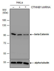
- Experimental details
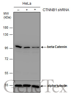
- Non-transfected (¡V) and transfected (+) HeLa whole cell extracts (30 ?g) were separated by 7.5% SDS-PAGE, and the membrane was blotted with beta Catenin antibody [N1N2-2], N-term (GTX101435) diluted at 1:1000. The HRP-conjugated anti-rabbit IgG antibody (GTX213110-01) was used to detect the primary antibody.
Supportive validation
- Submitted by
- GeneTex (provider)
- Main image

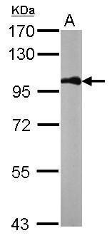
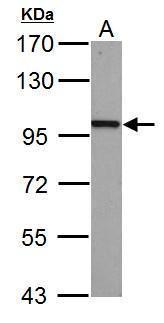
- Experimental details
- Sample (50 ?g of whole cell lysate) A: mouse brain 7.5% SDS PAGE GTX101435 diluted at 1:1000 The HRP-conjugated anti-rabbit IgG antibody (GTX213110-01) was used to detect the primary antibody.
- Submitted by
- GeneTex (provider)
- Main image

- Experimental details
- beta Catenin antibody [N1N2-2], N-term detects CTNNB1 protein by western blot analysis.A. 30 ?g PC-12 whole cell lysate/extract | 7.5% SDS-PAGEbeta Catenin antibody [N1N2-2], N-term (GTX101435) dilution: 1:1000 The HRP-conjugated anti-rabbit IgG antibody (GTX213110-01) was used to detect the primary antibody.
- Submitted by
- GeneTex (provider)
- Main image

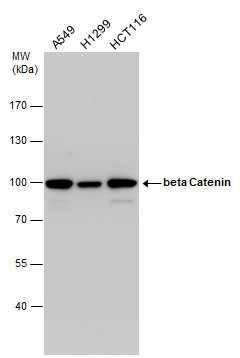
- Experimental details
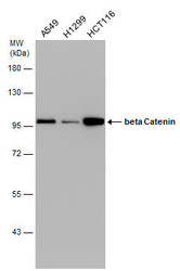
- beta Catenin antibody detects beta Catenin protein by western blot analysis. Various whole cell extracts (30 ?g) were separated by 7.5% SDS-PAGE, and the membrane was blotted with beta Catenin antibody (GTX101435) diluted by 1:1000.
- Validation comment
- WB
- Submitted by
- GeneTex (provider)
- Main image
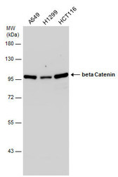
- Experimental details
- Various whole cell extracts (30 ?g) were separated by 7.5% SDS-PAGE, and the membrane was blotted with beta Catenin antibody [N1N2-2], N-term (GTX101435) diluted at 1:3000.

- Submitted by
- GeneTex (provider)
- Main image

- Experimental details
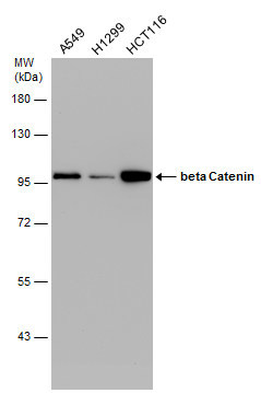
- Various whole cell extracts (30 ?g) were separated by 7.5% SDS-PAGE, and the membrane was blotted with beta Catenin antibody [N1N2-2], N-term (GTX101435) diluted at 1:10000.
- Submitted by
- GeneTex (provider)
- Main image
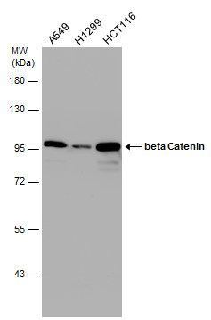
- Experimental details
- Various whole cell extracts (30 ?g) were separated by 7.5% SDS-PAGE, and the membrane was blotted with beta Catenin antibody [N1N2-2], N-term (GTX101435) diluted at 1:10000. The HRP-conjugated anti-rabbit IgG antibody (GTX213110-01) was used to detect the primary antibody.
- Submitted by
- GeneTex (provider)
- Main image

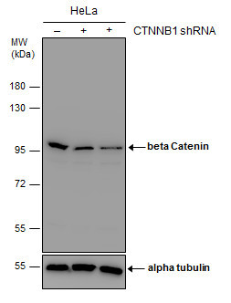
- Experimental details
- Non-transfected (¡V) and transfected (+) HeLa whole cell extracts (30 ?g) were separated by 7.5% SDS-PAGE, and the membrane was blotted with beta Catenin antibody [N1N2-2], N-term (GTX101435) diluted at 1:1000. The HRP-conjugated anti-rabbit IgG antibody (GTX213110-01) was used to detect the primary antibody.
Supportive validation
- Submitted by
- GeneTex (provider)
- Main image

- Experimental details
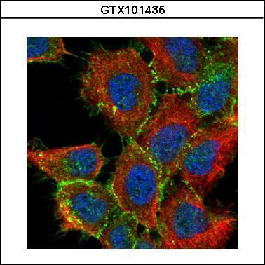
- Confocal immunofluorescence analysis (Olympus FV10i) of paraformaldehyde-fixed A431, using beta Catenin(GTX101435) antibody (Green) at 1:200 dilution. Alpha-tubulin filaments were labeled with GTX11304 (Red) at 1:2000.
- Submitted by
- GeneTex (provider)
- Main image

- Experimental details
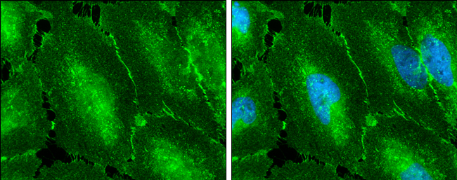
- beta Catenin antibody [N1N2-2], N-term detects beta Catenin protein at cell membrane by immunofluorescent analysis.Sample: HeLa cells were fixed in 4% paraformaldehyde at RT for 15 min.Green: beta Catenin protein stained by beta Catenin antibody [N1N2-2], N-term (GTX101435) diluted at 1:500.Blue: Hoechst 33342 staining.
- Submitted by
- GeneTex (provider)
- Main image
- Experimental details
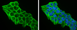
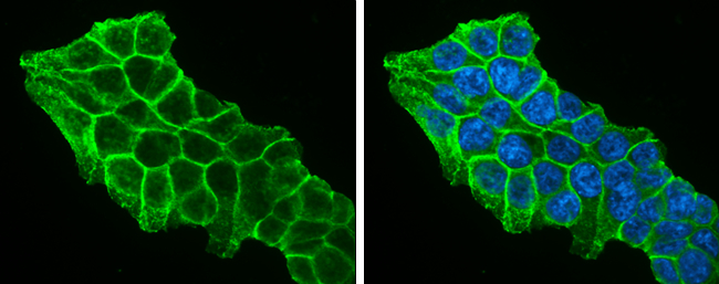
- beta Catenin antibody [N1N2-2], N-term detects beta Catenin protein at cell membrane by immunofluorescent analysis.Sample: HCT 116 cells were fixed in 4% paraformaldehyde at RT for 15 min.Green: beta Catenin protein stained by beta Catenin antibody [N1N2-2], N-term (GTX101435) diluted at 1:500.Blue: Hoechst 33342 staining.
Supportive validation
- Submitted by
- GeneTex (provider)
- Main image

- Experimental details
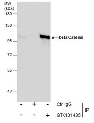
- beta Catenin antibody [N1N2-2], N-term immunoprecipitates CTNNB1 protein in IP experiments.IP samples: HeLa whole cell extractA. 30 ?g HeLa whole cell extractB. Control with 4 ?g of preimmune Rabbit IgGC. Immunoprecipitation of CTNNB1 protein by 4 ?g beta Catenin antibody [N1N2-2], N-term (GTX101435)5 % SDS-PAGEThe immunoprecipitated CTNNB1 protein was detected by beta Catenin antibody [N1N2-2], N-term (GTX101435) diluted at 1:1000.[EasyBlot anti-rabbit IgG (GTX221666-01) was used as a secondary reagent]
- Validation comment
- IP

- Submitted by
- GeneTex (provider)
- Main image
- Experimental details
- Immunoprecipitation of beta Catenin protein from HeLa whole cell extracts using 5 £gg of beta Catenin antibody (GTX101435).Western blot analysis was performed using beta Catenin antibody (GTX101435).EasyBlot anti-Rabbit IgG (GTX221666-01) was used as a secondary reagent.

Supportive validation
- Submitted by
- GeneTex (provider)
- Main image

- Experimental details
- beta Catenin antibody [N1N2-2], N-term detects beta Catenin protein at membrane on mouse skin by immunohistochemical analysis. Sample: Paraffin-embedded mouse skin. beta Catenin antibody [N1N2-2], N-term (GTX101435) dilution: 1:500.

- Submitted by
- GeneTex (provider)
- Main image

- Experimental details
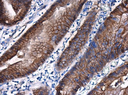
- beta Catenin antibody [N1N2-2], N-term detects beta Catenin protein at membrane on mouse colon by immunohistochemical analysis. Sample: Paraffin-embedded mouse colon. beta Catenin antibody [N1N2-2], N-term (GTX101435) dilution: 1:500.
- Submitted by
- GeneTex (provider)
- Main image

- Experimental details
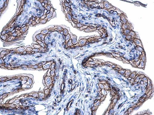
- beta Catenin antibody [N1N2-2], N-term detects beta Catenin protein at membrane on mouse urinary bladder by immunohistochemical analysis. Sample: Paraffin-embedded mouse urinary bladder. beta Catenin antibody [N1N2-2], N-term (GTX101435) diluted at 1:500.
- Submitted by
- GeneTex (provider)
- Main image

- Experimental details
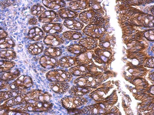
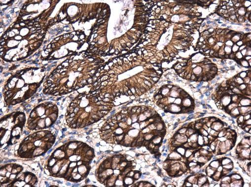
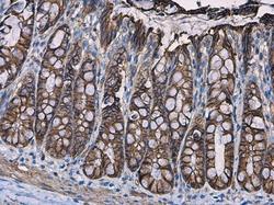
- beta Catenin antibody [N1N2-2], N-term detects beta Catenin protein at cell membrane and cytoplasm in rat colon by immunohistochemical analysis. Sample: Paraffin-embedded rat colon. beta Catenin antibody [N1N2-2], N-term (GTX101435) diluted at 1:500.
- Submitted by
- GeneTex (provider)
- Main image

- Experimental details
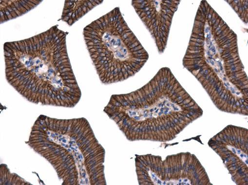
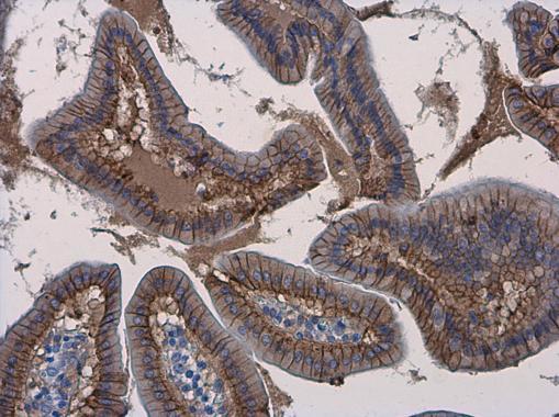
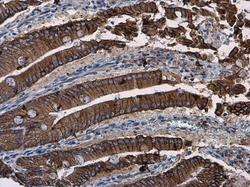
- beta Catenin antibody [N1N2-2], N-term detects beta Catenin protein at cell membrane and cytoplasm in mouse duodenum by immunohistochemical analysis. Sample: Paraffin-embedded mouse duodenum. beta Catenin antibody [N1N2-2], N-term (GTX101435) diluted at 1:500.
- Submitted by
- GeneTex (provider)
- Main image

- Experimental details
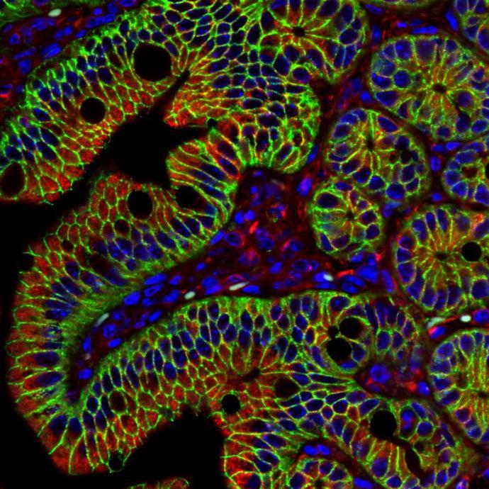
- beta Catenin antibody [N1N2-2] detects beta Catenin protein at cell membrane in mouse colon by immunohistochemical analysis. Sample: Paraffin-embedded mouse colon. Green: beta Catenin antibody [N1N2-2] (GTX101435) diluted at 1:500.Red: alpha Tubulin antibody [GT114] (GTX628802) diluted at 1:500. Blue: Hoechst 33342 staining.
- Submitted by
- GeneTex (provider)
- Main image

- Experimental details
- beta Catenin antibody [N1N2-2], N-term detects beta Catenin protein at cell membrane and cytoplasm in human esophagus by immunohistochemical analysis. Sample: Paraffin-embedded human esophagus. beta Catenin antibody [N1N2-2], N-term (GTX101435) diluted at 1:500.
- Submitted by
- GeneTex (provider)
- Main image

- Experimental details
- beta Catenin antibody [N1N2-2], N-term detects beta Catenin protein at cell membrane and cytoplasm in human cervix by immunohistochemical analysis. Sample: Paraffin-embedded human cervix. beta Catenin antibody [N1N2-2], N-term (GTX101435) diluted at 1:500.

- Submitted by
- GeneTex (provider)
- Main image

- Experimental details
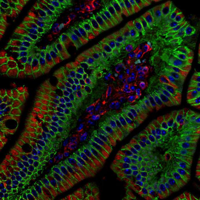
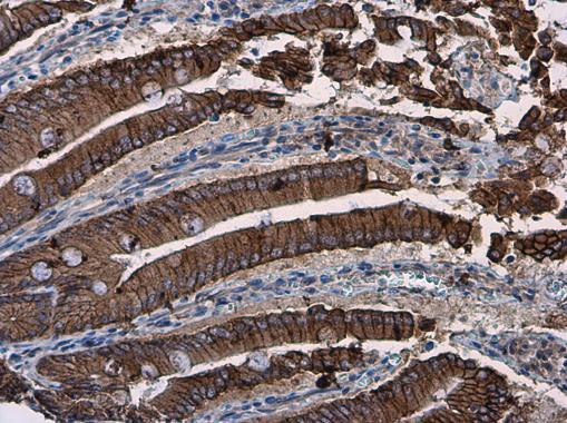
- beta Catenin antibody [N1N2-2], N-term detects beta Catenin protein at cell membrane and cytoplasm in mouse intestine by immunohistochemical analysis. Sample: Paraffin-embedded mouse intestine. beta Catenin antibody [N1N2-2], N-term (GTX101435) diluted at 1:500.

- Submitted by
- GeneTex (provider)
- Main image

- Experimental details
- beta Catenin antibody [N1N2-2], N-term detects beta Catenin protein at cell membrane and cytoplasm in mouse duodenum by immunohistochemical analysis. Sample: Paraffin-embedded mouse duodenum. beta Catenin antibody [N1N2-2], N-term (GTX101435) diluted at 1:500.
- Submitted by
- GeneTex (provider)
- Main image
- Experimental details
- beta Catenin antibody [N1N2-2], N-term detects beta Catenin protein at cell membrane and cytoplasm in rat colon by immunohistochemical analysis. Sample: Paraffin-embedded rat colon. beta Catenin antibody [N1N2-2], N-term (GTX101435) diluted at 1:500.

- Submitted by
- GeneTex (provider)
- Main image
- Experimental details
- beta Catenin antibody [N1N2-2], N-term detects beta Catenin protein at cell membrane and cytoplasm in rat duodenum by immunohistochemical analysis. Sample: Paraffin-embedded rat duodenum. beta Catenin antibody [N1N2-2], N-term (GTX101435) diluted at 1:500.
Supportive validation
- Submitted by
- GeneTex (provider)
- Main image
- Experimental details
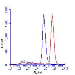
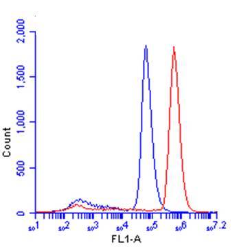
- beta Catenin antibody [N1N2-2], N-term (GTX101435) detects CTNNB1 protein by flow cytometry analysis. Sample: HeLa cell. Black: Unlabelled sample was used as a control. Red: beta Catenin antibody [N1N2-2], N-term (GTX101435) dilution: 1:50. Acquisition of 20,000 events were collected using a Dylight 488-conjugated secondary antibody for FACS analysis.
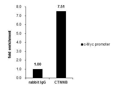
Supportive validation
- Submitted by
- GeneTex (provider)
- Main image
- Experimental details
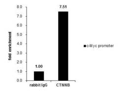
- Cross-linked ChIP was performed with HCT116 chromatin extract and 5 £gg of either control rabbit IgG or anti-beta Catenin antibody. The precipitated DNA was detected by PCR with primer set targeting to c-Myc promoter.