Explore
 Validate
 Learn
Learn Western blot
Antibody data
- Antibody Data
- Antigen structure
- References [0]
- Comments [0]
- Validations
- Western blot [2]
- Immunocytochemistry [2]
- Immunohistochemistry [8]
Submit
Validation data
Reference
Comment
Report error
- Product number
- AMAb91209 - Provider product page

- Provider
- Atlas Antibodies
- Proper citation
- Atlas Antibodies Cat#AMAb91209, RRID:AB_2665844
- Product name
- Anti-CTNNB1
- Antibody type
- Monoclonal
- Reactivity
- Human
- Host
- Mouse
- Conjugate
- Unconjugated
- Antigen sequence
SYLDSGIHSGATTTAPSLSGKGNPEEEDVDTSQVL
YEWEQGFSQSFTQEQVADIDGQYAMTRAQRVRAAM
FPETLDEGMQIPSTQFDAAH- Epitope
- Binds to an epitope located within the peptide sequence TSQVLYEWEQGFSQS as determined by overlapping synthetic peptides.
- Isotype
- IgG
- Antibody clone number
- CL3689
- Vial size
- 100 µl
- Storage
- Store at +4°C for short term storage. Long time storage is recommended at -20°C.
No comments: Submit comment
Supportive validation
- Submitted by
- Atlas Antibodies (provider)
- Main image
- Experimental details
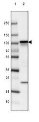
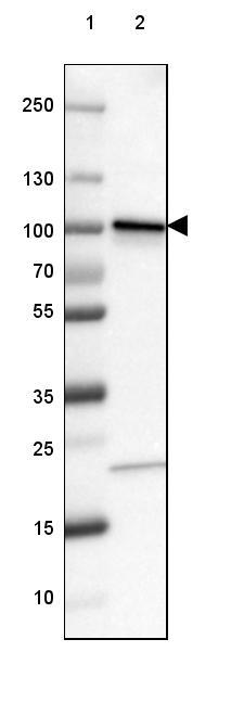
- Lane 1: Marker [kDa] 250, 130, 100, 70, 55, 35, 25, 15, 10Lane 2: Human cell line SK-MEL-30

- Submitted by
- Atlas Antibodies (provider)
- Main image

- Experimental details
- Lane 1: Marker [kDa] 250, 130, 100, 70, 55, 35, 25, 15, 10Lane 2: Human cell line A-549
Supportive validation
- Submitted by
- Atlas Antibodies (provider)
- Main image

- Experimental details
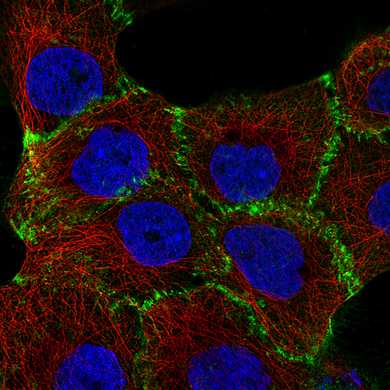
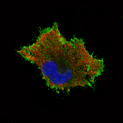
- Immunofluorescence staining of A-431 cells using the Anti-CTNNB1 monoclonal antibody, showing specific staining in the plasma membrane in green. Microtubule- and nuclear probes are visualized in red and blue, respectively (where available).
- Sample type
- HUMAN
- Submitted by
- Atlas Antibodies (provider)
- Main image
- Experimental details
- Immunofluorescence staining of U-251 cells using the Anti-CTNNB1 monoclonal antibody, showing specific staining in the plasma membrane in green. Microtubule- and nuclear probes are visualized in red and blue, respectively (where available).
- Sample type
- HUMAN

Supportive validation
- Submitted by
- Atlas Antibodies (provider)
- Main image
- Experimental details
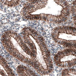
- Immunohistochemical staining of human endometrium shows strong membranous immunoreactivity in glandular epithelium.

- Submitted by
- Atlas Antibodies (provider)
- Main image

- Experimental details
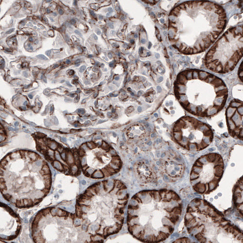
- Immunohistochemical staining of human kidney shows strong membranous positivity in renal tubules and glomerulus cells.
- Submitted by
- Atlas Antibodies (provider)
- Main image
- Experimental details
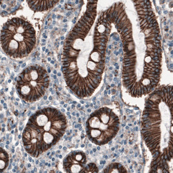
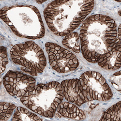
- Immunohistochemical staining of human duodenum shows strong membranous immunoreactivity in epithelial cells.

- Submitted by
- Atlas Antibodies (provider)
- Main image
- Experimental details
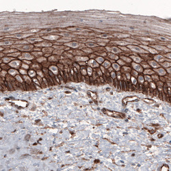
- Immunohistochemical staining of human cervix shows strong membranous positivity in epithelial cells.

- Submitted by
- Atlas Antibodies (provider)
- Main image

- Experimental details
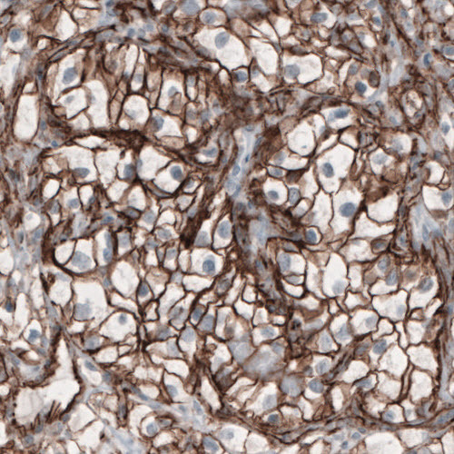
- Immunohistochemical staining of human breast cancer shows membranous immunoreactivity in tumor cells.
- Submitted by
- Atlas Antibodies (provider)
- Main image
- Experimental details
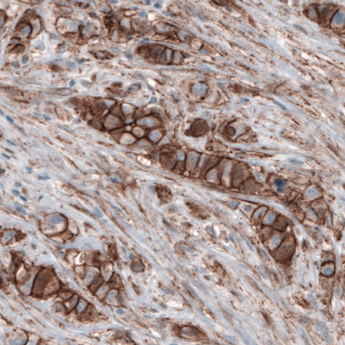
- Immunohistochemical staining of human prostate cancer shows membranous positivity in tumor cells.

- Submitted by
- Atlas Antibodies (provider)
- Main image

- Experimental details
- Immunohistochemical staining of human renal cancer shows membranous positivity in tumor cells.
- Submitted by
- Atlas Antibodies (provider)
- Main image
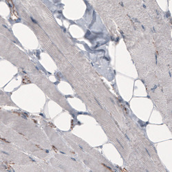
- Experimental details
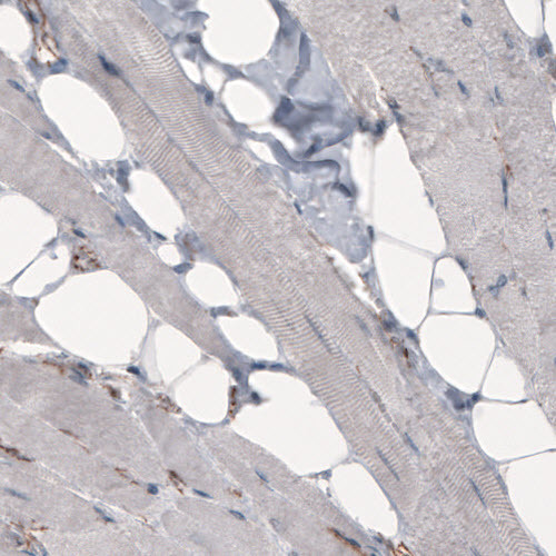
- Immunohistochemical staining of human skeletal muscle shows absence of immunoreactivity in muscle fibers (negative control).