 Explore
 Validate
 Learn
Learn Western blot
Antibody data
- Antibody Data
- Antigen structure
- References [2]
- Comments [0]
- Validations
- Western blot [7]
- Immunocytochemistry [4]
- Immunohistochemistry [12]
- Flow cytometry [1]
- Chromatin Immunoprecipitation [2]
- Other assay [2]
Submit
Validation data
Reference
Comment
Report error
- Product number
- PA5-77934 - Provider product page

- Provider
- Invitrogen Antibodies
- Product name
- beta Catenin Polyclonal Antibody
- Antibody type
- Polyclonal
- Antigen
- Recombinant full-length protein
- Description
- Positive Control: HeLa, A549, H1299, HCT116, PC-12, Mouse brain, human fibroblast, LX2
- Concentration
- 0.07 mg/mL
Submitted references Butyrate Inhibits Colorectal Cancer Cell Proliferation through Autophagy Degradation of β-Catenin Regardless of APC and β-Catenin Mutational Status.
FAT4 silencing promotes epithelial-to-mesenchymal transition and invasion via regulation of YAP and β-catenin activity in ovarian cancer.
Garavaglia B, Vallino L, Ferraresi A, Esposito A, Salwa A, Vidoni C, Gentilli S, Isidoro C
Biomedicines 2022 May 13;10(5)
Biomedicines 2022 May 13;10(5)
FAT4 silencing promotes epithelial-to-mesenchymal transition and invasion via regulation of YAP and β-catenin activity in ovarian cancer.
Malgundkar SH, Burney I, Al Moundhri M, Al Kalbani M, Lakhtakia R, Okamoto A, Tamimi Y
BMC cancer 2020 May 4;20(1):374
BMC cancer 2020 May 4;20(1):374
No comments: Submit comment
Supportive validation
- Submitted by
- Invitrogen Antibodies (provider)
- Main image

- Experimental details
- Western blot analysis of beta Catenin in non-transfected (-) and transfected (+) HeLa cells using 30 µg of protein. Samples were separated with 7.5% SDS-PAGE and incubated with beta Catenin polyclonal antibody (Product # PA5-77934) using a dilution of 1:1000 followed by HRP-conjugated anti-rabbit IgG.

- Submitted by
- Invitrogen Antibodies (provider)
- Main image
- Experimental details
- Western blot analysis of beta Catenin in mouse brain lysate using 50 µg of protein. Samples were separated with 7.5% SDS-PAGE and incubated with beta Catenin polyclonal antibody (Product # PA5-77934) using a dilution of 1:1000 followed by HRP-conjugated anti-rabbit IgG.

- Submitted by
- Invitrogen Antibodies (provider)
- Main image

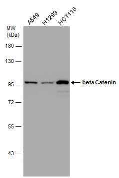
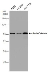
- Experimental details
- Western blot analysis of beta Catenin in whole cell lysate using 30 µg of protein. Samples were separated with 7.5% SDS-PAGE and incubated with beta Catenin polyclonal antibody (Product # PA5-77934) using a dilution of 1:10,000.

- Submitted by
- Invitrogen Antibodies (provider)
- Main image

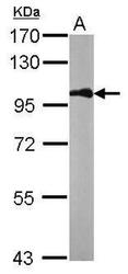
- Experimental details
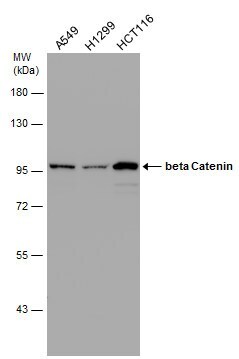
- Western Blot using beta Catenin Polyclonal Antibody (Product # PA5-77934). Various whole cell extracts (30 µg) were separated by 7.5% SDS-PAGE, and the membrane was blotted with beta Catenin Polyclonal Antibody (Product # PA5-77934) diluted at 1:10,000.
- Submitted by
- Invitrogen Antibodies (provider)
- Main image
- Experimental details
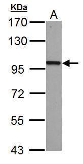
- Western Blot using beta Catenin Polyclonal Antibody (Product # PA5-77934). Sample (50 µg of whole cell lysate). Lane A: mouse brain. 7.5% SDS PAGE. beta Catenin Polyclonal Antibody (Product # PA5-77934) diluted at 1:1,000. The HRP-conjugated anti-rabbit IgG antibody was used to detect the primary antibody.

- Submitted by
- Invitrogen Antibodies (provider)
- Main image
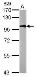
- Experimental details
- Western Blot using beta Catenin Polyclonal Antibody (Product # PA5-77934). Various whole cell extracts (30 µg) were separated by 7.5% SDS-PAGE, and the membrane was blotted with beta Catenin Polyclonal Antibody (Product # PA5-77934) diluted at 1:10,000.
- Submitted by
- Invitrogen Antibodies (provider)
- Main image

- Experimental details
- beta Catenin Polyclonal Antibody detects CTNNB1 protein by western blot analysis. A. 30 µg PC-12 whole cell lysate/extract .7.5% SDS-PAGE. Beta Catenin Polyclonal Antibody (Product # PA5-77934) dilution: 1:1,000. The HRP-conjugated anti-rabbit IgG antibody was used to detect the primary antibody.
Supportive validation
- Submitted by
- Invitrogen Antibodies (provider)
- Main image

- Experimental details
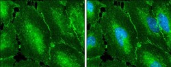
- Immunofluorescent analysis of beta Catenin in A431 cells. Samples were treated with paraformaldehyde and incubated with beta Catenin polyclonal antibody (Product # PA5-77934) using a dilution of 1:2000, followed by alpha tubulin (red) at a dilution of 1:2000.
- Submitted by
- Invitrogen Antibodies (provider)
- Main image

- Experimental details
- Immunocytochemistry-Immunofluorescence analysis of beta Catenin in paraformaldehyde-fixed A431 cells using beta Catenin Polyclonal Antibody (Product # PA5 77934) (Green) at a dilution of 1:200. Red: Alpha tubulin filaments.
- Submitted by
- Invitrogen Antibodies (provider)
- Main image
- Experimental details
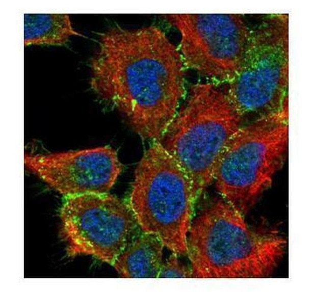
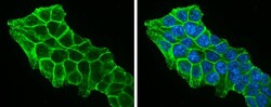
- beta Catenin Polyclonal Antibody detects beta Catenin protein at cell membrane by immunofluorescent analysis. Sample: HCT 116 cells were fixed in 4% paraformaldehyde at RT for 15 min. Green: beta Catenin protein stained by beta Catenin Polyclonal Antibody (Product # PA5-77934) diluted at 1:500. Blue: Hoechst 33342 staining.

- Submitted by
- Invitrogen Antibodies (provider)
- Main image
- Experimental details
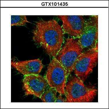
- beta Catenin Polyclonal Antibody detects beta Catenin protein at cell membrane by immunofluorescent analysis. Sample: HeLa cells were fixed in 4% paraformaldehyde at RT for 15 min. Green: beta Catenin protein stained by beta Catenin Polyclonal Antibody (Product # PA5-77934) diluted at 1:500. Blue: Hoechst 33342 staining.
Supportive validation
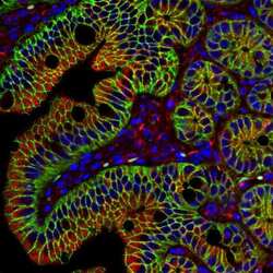
- Submitted by
- Invitrogen Antibodies (provider)
- Main image

- Experimental details
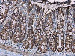
- beta Catenin antibody [N1N2-2] detects beta Catenin protein at cell membrane in mouse colon by immunohistochemical analysis. Sample: Paraffin-embedded mouse colon. Green: beta Catenin antibody [N1N2-2] (Product # PA5-77934) diluted at 1:500. Red: alpha Tubulin antibody [GT114] (Product # MA5-31466) diluted at 1:500. Blue: Hoechst 33342 staining. Antigen Retrieval: Citrate buffer, pH 6.0, 15 min.
- Submitted by
- Invitrogen Antibodies (provider)
- Main image

- Experimental details
- beta Catenin Polyclonal Antibody detects beta Catenin protein at membrane on mouse colon by immunohistochemical analysis. Sample: Paraffin-embedded mouse colon. Beta Catenin Polyclonal Antibody (Product # PA5-77934) dilution: 1:500. Antigen Retrieval: EDTA based buffer, pH 8.0, 15 min.

- Submitted by
- Invitrogen Antibodies (provider)
- Main image

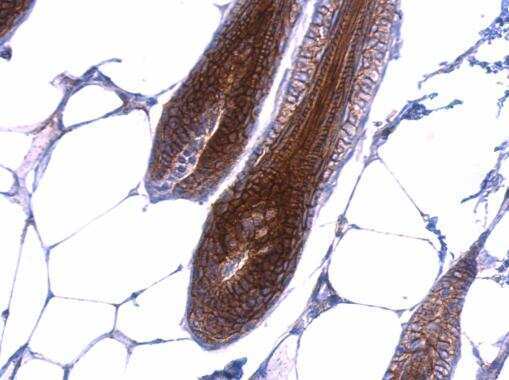
- Experimental details
- beta Catenin Polyclonal Antibody detects beta Catenin protein at membrane on mouse skin by immunohistochemical analysis. Sample: Paraffin-embedded mouse skin. Beta Catenin Polyclonal Antibody (Product # PA5-77934) dilution: 1:500. Antigen Retrieval: EDTA based buffer, pH 8.0, 15 min.
- Submitted by
- Invitrogen Antibodies (provider)
- Main image
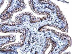
- Experimental details
- beta Catenin Polyclonal Antibody detects beta Catenin protein at membrane on mouse urinary bladder by immunohistochemical analysis. Sample: Paraffin-embedded mouse urinary bladder. Beta Catenin Polyclonal Antibody (Product # PA5-77934) diluted at 1:500. Antigen Retrieval: EDTA based buffer, pH 8.0, 15 min.

- Submitted by
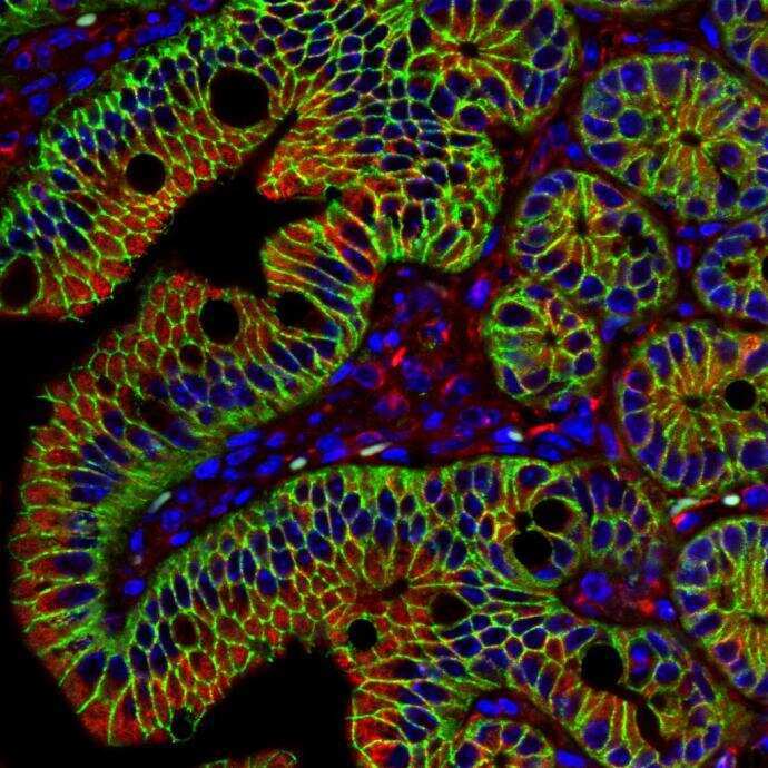
- Invitrogen Antibodies (provider)
- Main image
- Experimental details
- beta Catenin antibody [N1N2-2] detects beta Catenin protein at cell membrane in mouse colon by immunohistochemical analysis. Sample: Paraffin-embedded mouse colon. Green: beta Catenin antibody [N1N2-2] (Product # PA5-77934) diluted at 1:500. Red: alpha Tubulin antibody [GT114] (Product # MA5-31466) diluted at 1:500. Blue: Hoechst 33342 staining. Antigen Retrieval: Citrate buffer, pH 6.0, 15 min.
- Submitted by
- Invitrogen Antibodies (provider)
- Main image
- Experimental details
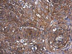
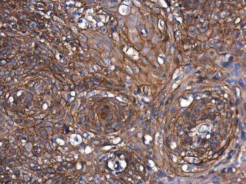
- beta Catenin Polyclonal Antibody detects beta Catenin protein at cell membrane and cytoplasm in human cervix by immunohistochemical analysis. Sample: Paraffin-embedded human cervix. Beta Catenin Polyclonal Antibody (Product # PA5-77934) diluted at 1:500. Antigen Retrieval: Citrate buffer, pH 6.0, 15 min.
- Submitted by
- Invitrogen Antibodies (provider)
- Main image
- Experimental details
- beta Catenin Polyclonal Antibody detects beta Catenin protein at cell membrane and cytoplasm in human esophagus by immunohistochemical analysis. Sample: Paraffin-embedded human esophagus. Beta Catenin Polyclonal Antibody (Product # PA5-77934) diluted at 1:500. Antigen Retrieval: Citrate buffer, pH 6.0, 15 min.
- Submitted by
- Invitrogen Antibodies (provider)
- Main image
- Experimental details
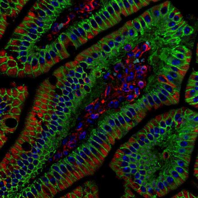
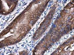
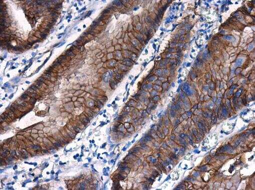
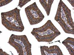
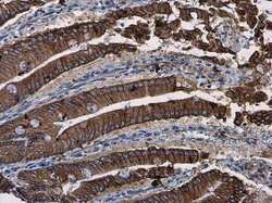
- beta Catenin Polyclonal Antibody detects beta Catenin protein at cell membrane and cytoplasm in mouse duodenum by immunohistochemical analysis. Sample: Paraffin-embedded mouse duodenum. Beta Catenin Polyclonal Antibody (Product # PA5-77934) diluted at 1:500. Antigen Retrieval: Citrate buffer, pH 6.0, 15 min.
- Submitted by
- Invitrogen Antibodies (provider)
- Main image
- Experimental details
- beta Catenin Polyclonal Antibody detects beta Catenin protein at cell membrane and cytoplasm in mouse duodenum by immunohistochemical analysis. Sample: Paraffin-embedded mouse duodenum. Beta Catenin Polyclonal Antibody (Product # PA5-77934) diluted at 1:500. Antigen Retrieval: Citrate buffer, pH 6.0, 15 min.

- Submitted by
- Invitrogen Antibodies (provider)
- Main image
- Experimental details
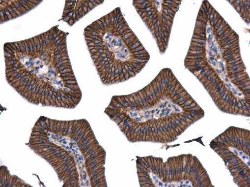
- beta Catenin Polyclonal Antibody detects beta Catenin protein at cell membrane and cytoplasm in mouse intestine by immunohistochemical analysis. Sample: Paraffin-embedded mouse intestine. Beta Catenin Polyclonal Antibody (Product # PA5-77934) diluted at 1:500. Antigen Retrieval: Citrate buffer, pH 6.0, 15 min.

- Submitted by
- Invitrogen Antibodies (provider)
- Main image
- Experimental details
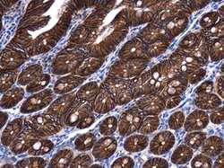
- beta Catenin Polyclonal Antibody detects beta Catenin protein at cell membrane and cytoplasm in rat colon by immunohistochemical analysis. Sample: Paraffin-embedded rat colon. Beta Catenin Polyclonal Antibody (Product # PA5-77934) diluted at 1:500. Antigen Retrieval: Citrate buffer, pH 6.0, 15 min.

- Submitted by
- Invitrogen Antibodies (provider)
- Main image
- Experimental details
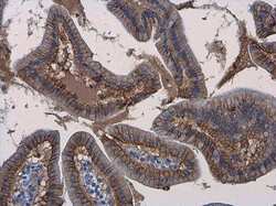
- beta Catenin Polyclonal Antibody detects beta Catenin protein at cell membrane and cytoplasm in rat duodenum by immunohistochemical analysis. Sample: Paraffin-embedded rat duodenum. Beta Catenin Polyclonal Antibody (Product # PA5-77934) diluted at 1:500. Antigen Retrieval: Citrate buffer, pH 6.0, 15 min.

Supportive validation
- Submitted by
- Invitrogen Antibodies (provider)
- Main image
- Experimental details
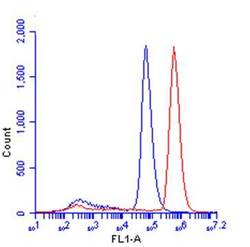
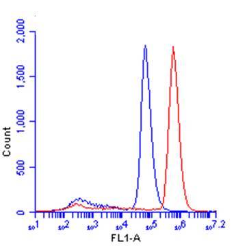
- beta Catenin Polyclonal Antibody (Product # PA5-77934) detects CTNNB1 protein by flow cytometry analysis. Sample: HeLa cell. Black: Unlabelled sample was used as a control. Red: beta Catenin Polyclonal Antibody (Product # PA5-77934) dilution: 1:50. Acquisition of 20,000 events were collected for Flow cytometry analysis analysis.
Supportive validation
- Submitted by
- Invitrogen Antibodies (provider)
- Main image

- Experimental details
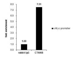
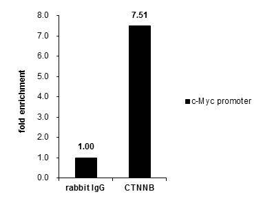
- Chromatin immunoprecipitation analysis of beta Catenin in HCT-116 chromatin extract using 5 µg of sample. Immunoprecipitation was performed using beta Catenin polyclonal antibody (Product # PA5-77934). Analysis of the precipitated DNA was done by PCR using primer targeting c-Myc promoter.

- Submitted by
- Invitrogen Antibodies (provider)
- Main image
- Experimental details
- Cross-linked ChIP was performed with HCT116 chromatin extract and 5 µg of either control rabbit IgG or beta Catenin Polyclonal Antibody (Product # PA5-77934). The precipitated DNA was detected by PCR with primer set targeting to c-Myc promoter.
Supportive validation
- Submitted by
- Invitrogen Antibodies (provider)
- Main image
- Experimental details
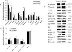
- Fig. 4 Regulatory effects of FAT4 on the expression of proteins involved in EMT, Hippo, Wnt-beta-catenin, apoptotic, and retinoblastoma pathways by Western blotting. a . Relative expression variation of proteins. The expression of Vimentin ( p = 0.0001), YAP ( p = 0.0018), beta-catenin ( p = 0.001), Bcl2 ( p = 0.0001), cyclin D1 ( p = 0.0017) and cdk4 ( p = 0.0025) was higher in FAT4 siRNA treated cells as compared to control. beta-actin was used as an internal control. b . Western blot demonstrating bands for protein expression in control and FAT4 knocked cells. Full-length blots are presented in Supplementary figure S 1 . c . The ratio of phosphorylated to total YAP, GSK-3beta, beta-catenin, and retinoblastoma proteins following FAT4 repression. The pYAP/ YAP ratio was lower in FAT4 siRNA treated cells as compared to the control ( p = 0.0286). Similarly, pGSK-3-beta/GSK-3-beta ratio, and pbeta-catenin/beta-catenin ratio was lower in FAT4 siRNA treated cells ( p = 0.018, and p = 0.001 respectively) as compared to control. There was no significant difference in pRb/Rb ratio between FAT4 siRNA treated cells and control. MCAS cells treated with scrambled siRNA was used as control. Data represent mean and standard deviation from at least three independent experiments performed in triplicates

- Submitted by
- Invitrogen Antibodies (provider)
- Main image
- Experimental details
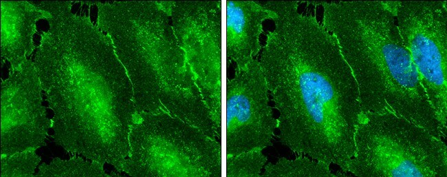
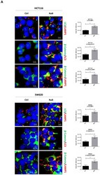
- Sodium butyrate induces beta-Catenin sequestration in the autophagy-lysosomal system. ( A ) HCT116 and SW620 cells adhering to coverslips were treated as indicated for 48 h, thereafter cells were fixed and double-stained for LC3 (green)/LAMP1 (red), beta-Catenin (green)/LC3 (red), or for beta-Catenin (green)/LAMP1 (red). Nuclei were stained with DAPI. The images were acquired by fluorescence microscope (scale bar = 8.5 mum, magnification = 63x). The images shown are representative of various fields for each condition. Bars in the graph indicate the average yellow fluorescence integrity density. Data are from three different images for each condition. The graphs represent the yellow integrity density +- SD (significance was considered as follow: * p < 0.05, ** p < 0.01). ( B ) A parallel set of cultures in petri dishes was processed for co-immunoprecipitation analysis of LC3 interacting protein. LC3 and beta-Catenin were revealed by Western blotting. The experiment was performed three times with similar results. Densitometric data are shown in the panel. The graphs represent the average +- SD (significance was considered as follow: ** p < 0.01, *** p < 0.001).
