Explore
 Validate
 Learn
Learn Western blot
Antibody data
- Antibody Data
- Antigen structure
- References [2]
- Comments [0]
- Validations
- Western blot [4]
- Immunocytochemistry [4]
- Immunohistochemistry [2]
- Flow cytometry [1]
- Other assay [1]
Submit
Validation data
Reference
Comment
Report error
- Product number
- MA5-32118 - Provider product page

- Provider
- Invitrogen Antibodies
- Product name
- Cytokeratin 8 Recombinant Rabbit Monoclonal Antibody (SU0338)
- Antibody type
- Monoclonal
- Antigen
- Synthetic peptide
- Reactivity
- Human, Mouse
- Host
- Rabbit
- Isotype
- IgG
- Antibody clone number
- SU0338
- Vial size
- 100 µL
- Concentration
- 1 mg/mL
- Storage
- Store at 4°C short term. For long term storage, store at -20°C, avoiding freeze/thaw cycles.
Submitted references High throughput, label-free isolation of circulating tumor cell clusters in meshed microwells.
A FACS-Free Purification Method to Study Estrogen Signaling, Organoid Formation, and Metabolic Reprogramming in Mammary Epithelial Cells.
Boya M, Ozkaya-Ahmadov T, Swain BE, Chu CH, Asmare N, Civelekoglu O, Liu R, Lee D, Tobia S, Biliya S, McDonald LD, Nazha B, Kucuk O, Sanda MG, Benigno BB, Moreno CS, Bilen MA, McDonald JF, Sarioglu AF
Nature communications 2022 Jun 13;13(1):3385
Nature communications 2022 Jun 13;13(1):3385
A FACS-Free Purification Method to Study Estrogen Signaling, Organoid Formation, and Metabolic Reprogramming in Mammary Epithelial Cells.
Lacouture A, Jobin C, Weidmann C, Berthiaume L, Bastien D, Laverdière I, Pelletier M, Audet-Walsh É
Frontiers in endocrinology 2021;12:672466
Frontiers in endocrinology 2021;12:672466
No comments: Submit comment
Supportive validation
- Submitted by
- Invitrogen Antibodies (provider)
- Main image
- Experimental details
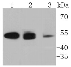
- Western blot analysis of Cytokeratin 8 in different lysates using a Monoclonal antibody (Product #MA5-32118) at a dilution of 1:1,000. Positive control: Lane 1: Hela, Lane 2: MCF-7, Lane 3: A431.
- Submitted by
- Invitrogen Antibodies (provider)
- Main image

- Experimental details
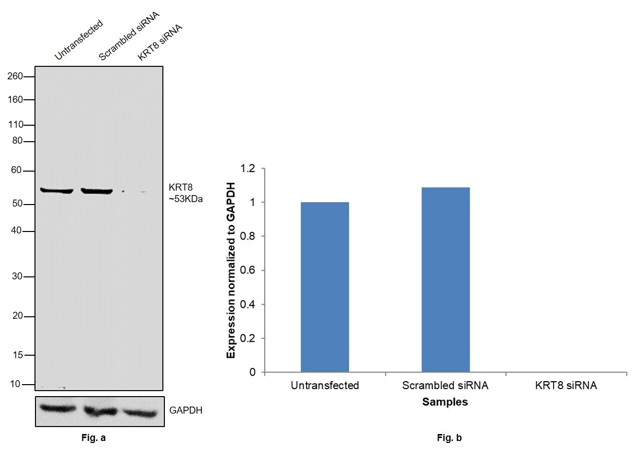
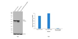
- Knockdown of KRT8 was achieved by transfecting Caco-2 with KRT8 specific siRNAs (Silencer® select Product # S7969, S7970). Western blot analysis (Fig. a) was performed using Whole cell extracts from the KRT8 knockdown cells (lane 3), non-targeting scrambled siRNA transfected cells (lane 2) and untransfected cells (lane 1). The blot was probed with Cytokeratin 8 Recombinant Rabbit Monoclonal Antibody (SU0338) (Product # MA5-32118, 1:1000 dilution) and Goat anti-Rabbit IgG (H+L) Superclonal™ Recombinant Secondary Antibody, HRP (Product # A27036, 1:4000). Densitometric analysis of this western blot is shown in histogram (Fig. b). Decrease in signal upon siRNA mediated knock down confirms that antibody is specific to KRT8.
- Submitted by
- Invitrogen Antibodies (provider)
- Main image
- Experimental details
- CRISPR-Cas9 mediated genome editing ofKRT8 (as confirmed by next generation sequencing) was achieved by using LentiArray™ Lentiviral sgRNA (Product # A32042, Assay ID CRISPR745510_LV) and LentiArray Cas9 Lentivirus (Product # A32064). Western blot analysis of KRT8 was performed by loading 30 µg of HeLa wild type (Lane 1), HeLa Cas9 (Lane 2) and HeLa Cas9 transduced with KRT8 Lentiviral sgRNA (Lane 3) whole cell extracts . The samples were electrophoresed using NuPAGE™ Novex™ 4-12% Bis-Tris Protein Gel (Product # NP0322BOX). Resolved proteins were then transferred onto a nitrocellulose membrane (Product # IB23001) by iBlot® 2 Dry Blotting System (Product # IB21001). The blot was probed with Cytokeratin 8 Recombinant Rabbit Monoclonal Antibody (SU0338) (Product # MA5-32118, 1:5000 dilution) and Goat anti-Rabbit IgG (H+L) Superclonal™ Recombinant Secondary Antibody, HRP (Product # A27036, 1:20,000 dilution) using the iBright™ FL1500 (Product # A44115). Chemiluminescent detection was performed using SuperSignal™ West Dura Extended Duration Substrate (Product # 34076). A reduced signal in sgRNA transduced cells using the LentiArray™ CRISPR product line confirms that antibody is specific toKRT8.

- Submitted by
- Invitrogen Antibodies (provider)
- Main image
- Experimental details
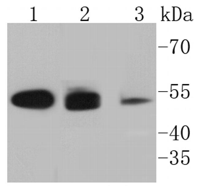
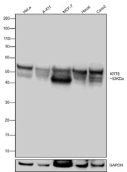
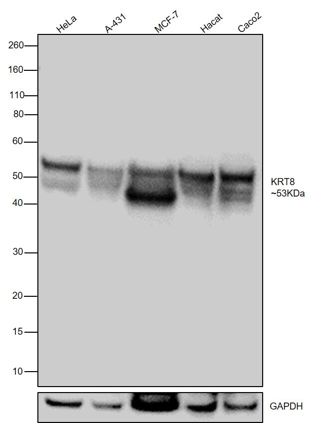
- Western blot was performed using Anti-Cytokeratin 8 Recombinant Rabbit Monoclonal Antibody (SU0338) (Product # MA5-32118) and a 53 kDa band corresponding to KRT8 was observed across the cell lines tested. Whole cell extracts (30 µg lysate) of HeLa (Lane 1), A-431 (Lane 2), MCF7 (Lane 3), HaCaT (Lane 4), Caco-2 (Lane 5) were electrophoresed using NuPAGE™ 4-12% Bis-Tris Protein Gel (Product # NP0322BOX). Resolved proteins were then transferred onto a nitrocellulose membrane (Product # IB23001) by iBlot® 2 Dry Blotting System (Product # IB21001). The blot was probed with the primary antibody (1:1000) and detected by chemiluminescence with Goat anti-Rabbit IgG (H+L) Superclonal™ Recombinant Secondary Antibody, HRP (Product # A27036,1:20,000) using the iBright™ FL1500 Imaging System (Product # A44115). Chemiluminescent detection was performed using SuperSignal™ West Pico PLUS Chemiluminescent Substrate (Product # 34580).
Supportive validation
- Submitted by
- Invitrogen Antibodies (provider)
- Main image

- Experimental details
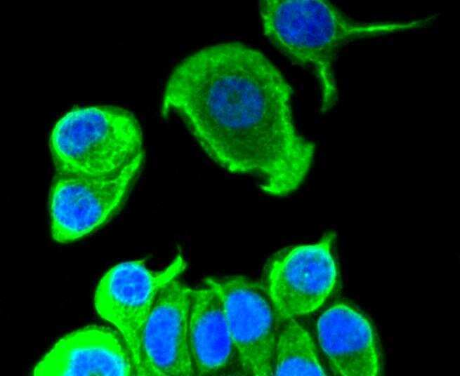
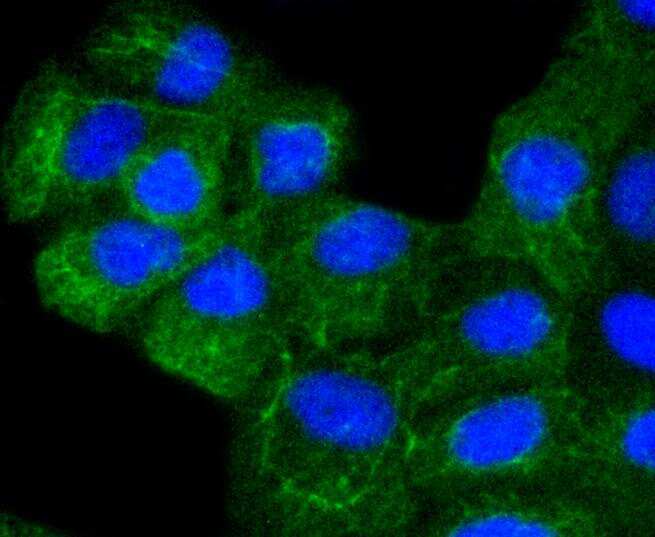
- Immunocytochemical analysis of Cytokeratin 8 in MCF-7 cells using a Cytokeratin 8 Monoclonal antibody (Product # MA5-32118) as seen in green. The nuclear counter stain is DAPI (blue). Cells were fixed in paraformaldehyde, permeabilised with 0.25% Triton X100/PBS.
- Submitted by
- Invitrogen Antibodies (provider)
- Main image

- Experimental details
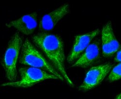
- Immunocytochemical analysis of Cytokeratin 8 in A431 cells using a Cytokeratin 8 Monoclonal antibody (Product # MA5-32118) as seen in green. The nuclear counter stain is DAPI (blue). Cells were fixed in paraformaldehyde, permeabilised with 0.25% Triton X100/PBS.
- Submitted by
- Invitrogen Antibodies (provider)
- Main image
- Experimental details
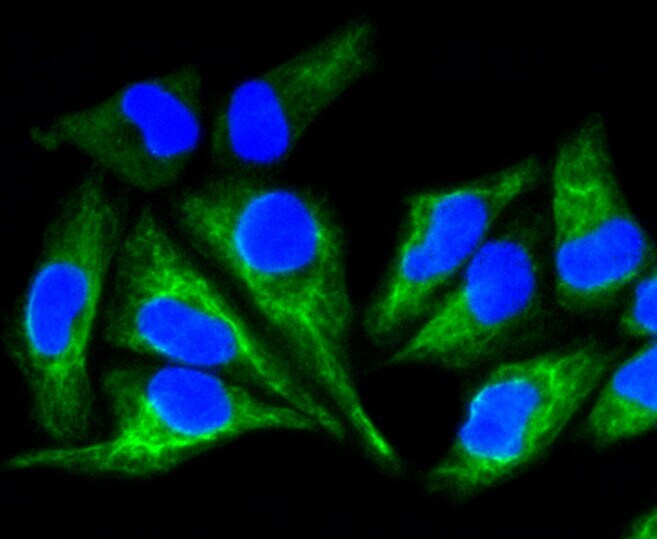
- Immunocytochemical analysis of Cytokeratin 8 in Hela cells using a Cytokeratin 8 Monoclonal antibody (Product # MA5-32118) as seen in green. The nuclear counter stain is DAPI (blue). Cells were fixed in paraformaldehyde, permeabilised with 0.25% Triton X100/PBS.
- Submitted by
- Invitrogen Antibodies (provider)
- Main image
- Experimental details
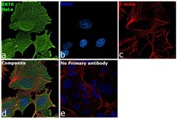
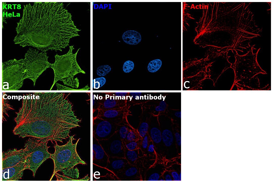
- Immunofluorescence analysis of KRT8 was performed using 70% confluent log phase HeLa cells. The cells were fixed with 4% paraformaldehyde for 10 minutes, permeabilized with 0.1% Triton™ X-100 for 15 minutes, and blocked with 2% BSA for 1 hour at room temperature. The cells were labeled with Cytokeratin 8 Recombinant Rabbit Monoclonal Antibody (SU0338) (Product # MA5-32118) at 1:500 in 0.1% BSA, incubated at 4 degree celsius overnight and then labeled with Goat anti-Rabbit IgG (H+L) Highly Cross-Adsorbed Secondary Antibody, Alexa Fluor Plus 488 (Product # A32731, 1:2000), for 45 minutes at room temperature (Panel a: Green). Nuclei (Panel b:Blue) were stained with ProLong™ Diamond Antifade Mountant with DAPI (Product # P36962). F-actin (Panel c: Red) was stained with Rhodamine Phalloidin (Product # R415, 1:300). Panel d represents the merged image showing Intermediate filaments localization. Panel e represents control cells with no primary antibody to assess background. The images were captured at 60X magnification.
Supportive validation
- Submitted by
- Invitrogen Antibodies (provider)
- Main image
- Experimental details
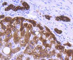
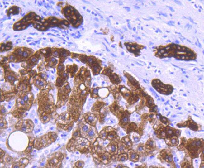
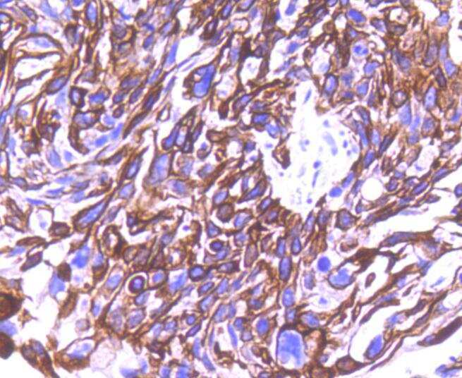
- Immunohistochemical analysis of Cytokeratin 8 of paraffin-embedded Human liver tissue using a Cytokeratin 8 Monoclonal antibody (Product #MA5-32118). Counter stained with hematoxylin.
- Submitted by
- Invitrogen Antibodies (provider)
- Main image

- Experimental details
- Immunohistochemical analysis of Cytokeratin 8 of paraffin-embedded Human liver tissue using a Cytokeratin 8 Monoclonal antibody (Product #MA5-32118). Counter stained with hematoxylin.
Supportive validation
- Submitted by
- Invitrogen Antibodies (provider)
- Main image
- Experimental details
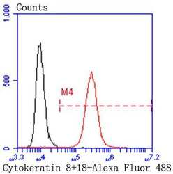
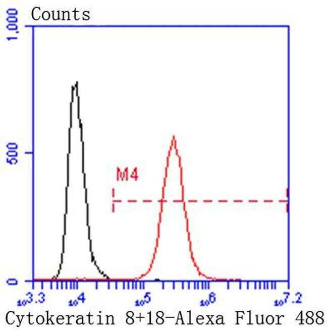
- Flow Cytometric analysis of Cytokeratin 8 in Hela cells using a Cytokeratin 8 Monoclonal Antibody (Product # MA5-32118) at a dilution of 1:50, as seen in red compared with an unlabelled control (cells without incubation with primary antibody; black). Alexa Fluor 488-conjugated goat anti rabbit IgG was used as the secondary antibody.
Supportive validation
- Submitted by
- Invitrogen Antibodies (provider)
- Main image

- Experimental details
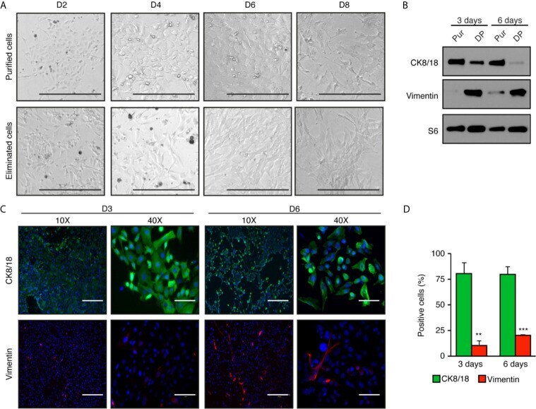
- Figure 2 High enrichment of mammary epithelial cell for primary culture in two-dimensions. (A) Brightfield images of mammary epithelial cells obtained after purification compared with the cells eliminated during the purification process. Scale bars = 300 mum. (B) Western blot analysis of purified cells normally discarded with differential plating (DP) compared to purified epithelial cells (Pur). Protein expression of the cytokeratin 8 and 18 (CK8/18), a marker of epithelial cells, and vimentin, a marker of fibroblasts, after three and six days in two-dimensional culture. S6 was used as the loading control. (C) Immunofluorescence showing the expression of CK8/18 (green) and vimentin (red) at three or six days in two-dimensional culture. Nuclei were stained with DAPI (blue). Scale bars = 300 mum and 75 mum. (D) Ratios of positive cells for CK8/18 or vimentin per the total number of cells (counts of nuclei) in percentage. Data are shown as mean +- SEM of one representative experiment (n = 6 images per condition). ** p < 0.01; *** p < 0.001.