Explore
 Validate
 Learn
Learn Western blot
Antibody data
- Antibody Data
- Antigen structure
- References [0]
- Comments [0]
- Validations
- Western blot [2]
- Immunohistochemistry [4]
- Flow cytometry [1]
Submit
Validation data
Reference
Comment
Report error
- Product number
- MA5-34674 - Provider product page

- Provider
- Invitrogen Antibodies
- Product name
- Podocin Recombinant Rabbit Monoclonal Antibody (JB51-33)
- Antibody type
- Monoclonal
- Antigen
- Synthetic peptide
- Description
- Positive Control: Rat kidney tissue, human kidney tissue, mouse kidney tissue, 293T.
- Reactivity
- Human, Mouse
- Host
- Rabbit
- Isotype
- IgG
- Antibody clone number
- JB51-33
- Vial size
- 100 µL
- Concentration
- 1 mg/mL
- Storage
- -20° C, Avoid Freeze/Thaw Cycles, store in dark
No comments: Submit comment
Supportive validation
- Submitted by
- Invitrogen Antibodies (provider)
- Main image

- Experimental details
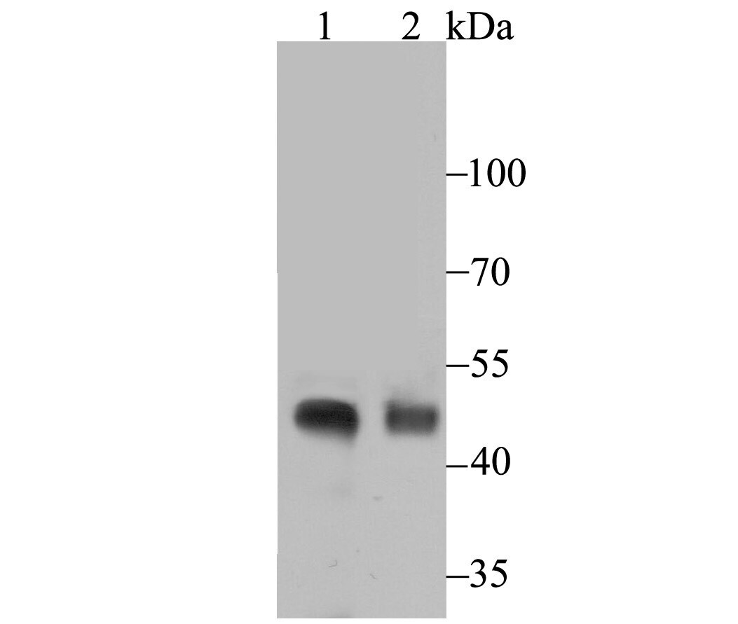
- Western blot analysis of NPHS2 in rat kidney (1) and human kidney (2) tissue lysate. Samples were incubated with NPHS2 monoclonal antibody (Product # MA5-34674), at a dilution of 1:500.
- Submitted by
- Invitrogen Antibodies (provider)
- Main image
- Experimental details
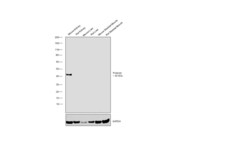
- Western blot was performed using Anti-Podocin Recombinant Rabbit Monoclonal Antibody (JB51-33) (Product # MA5-34674) and a ~42 kDa band corresponding to Nphs2 was observed only in mouse kidney tissue. Whole cell extracts (30 µg lysate) of Mouse Kidney (Lane 1), Rat Kidney (Lane 2), Mouse Liver (Lane 3), Rat Liver (Lane 4), Mouse Skeletal Muscle (Lane 5), Rat Skeletal Muscle (Lane 6) were electrophoresed using NuPAGE™ 4-12% Bis-Tris Protein Gel (Product # NP0322BOX). Resolved proteins were then transferred onto a nitrocellulose membrane (Product # IB23002) by iBlot® 2 Dry Blotting System (Product # IB21001). The blot was probed with the primary antibody (1:1000) and detected by chemiluminescence with Goat anti-Rabbit IgG (H+L) Superclonal™ Recombinant Secondary Antibody, HRP (Product # A27036,1:20000) using the iBright™ FL1500 Imaging System (Product # A44115). Chemiluminescent detection was performed using SuperSignal™ West Pico PLUS Chemiluminescent Substrate (Product # 34580).

Supportive validation
- Submitted by
- Invitrogen Antibodies (provider)
- Main image
- Experimental details
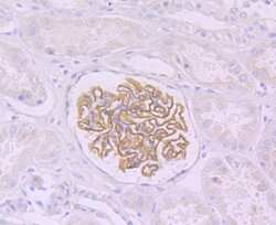
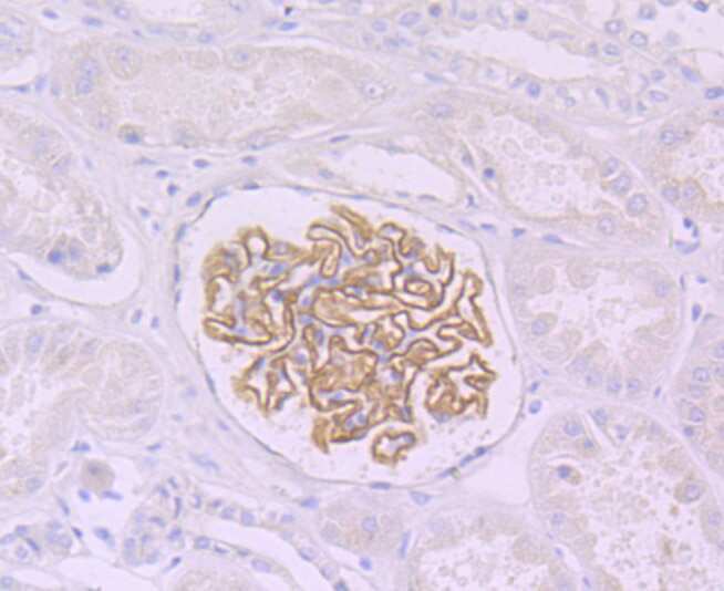
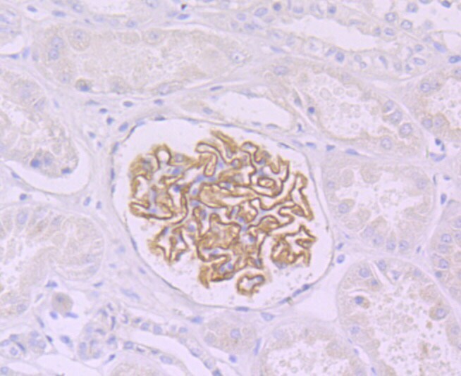
- Immunohistochemistry analysis of NPHS2 in paraffin-embedded human kidney tissue. Samples were incubated with NPHS2 monoclonal antibody (Product # MA5-34674), and followed by hematoxylin.
- Submitted by
- Invitrogen Antibodies (provider)
- Main image
- Experimental details
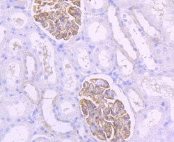
- Immunohistochemistry analysis of NPHS2 in paraffin-embedded rat kidney tissue. Samples were incubated with NPHS2 monoclonal antibody (Product # MA5-34674), and followed by hematoxylin.

- Submitted by
- Invitrogen Antibodies (provider)
- Main image

- Experimental details
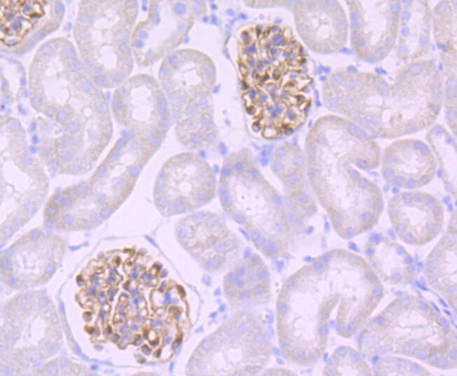
- Immunohistochemistry analysis of NPHS2 in paraffin-embedded mouse kidney tissue. Samples were incubated with NPHS2 monoclonal antibody (Product # MA5-34674), and followed by hematoxylin.
- Submitted by
- Invitrogen Antibodies (provider)
- Main image

- Experimental details
- Immunohistochemistry analysis of NPHS2 in paraffin-embedded human kidney tissue. Samples were incubated with NPHS2 monoclonal antibody (Product # MA5-34674), and followed by hematoxylin.
Supportive validation
- Submitted by
- Invitrogen Antibodies (provider)
- Main image
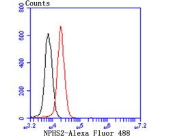
- Experimental details
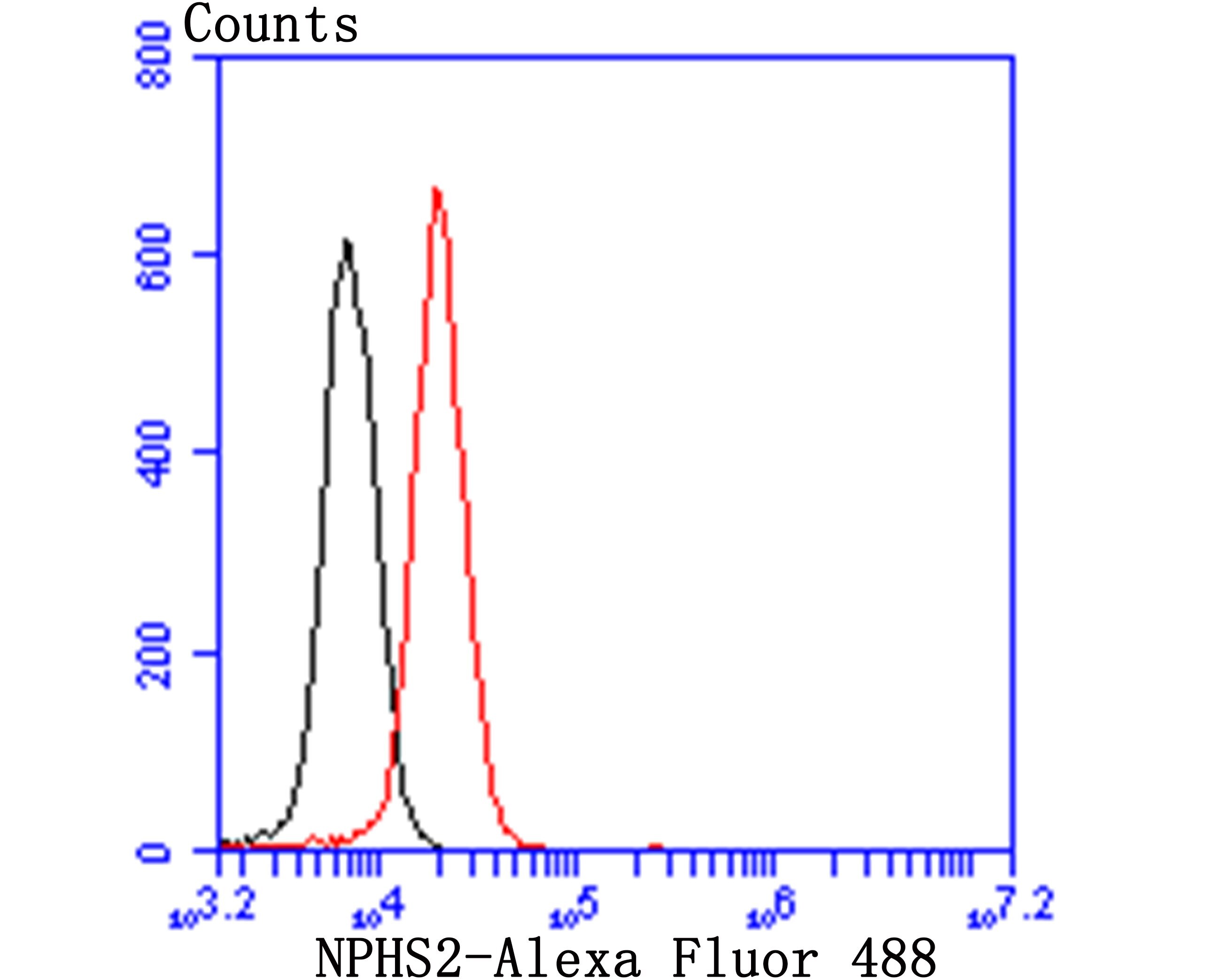
- Flow cytometry of NPHS2 in 293T cells (red) compared with an unlabelled control (cells without incubation with primary antibody; black). Samples were incubated with NPHS2 monoclonal antibody (Product # MA5-34674) at a dilution of 1:50, followed by Alexa Fluor 488-conjugated goat anti-rabbit IgG.