 Explore
 Validate
 Learn
Learn Western blot
 Immunohistochemistry
Antibody data
- Antibody Data
- Antigen structure
- References [0]
- Comments [0]
- Validations
- Immunohistochemistry [3]
Submit
Validation data
Reference
Comment
Report error
- Product number
- NB120-2737 - Provider product page

- Provider
- Novus Biologicals
- Proper citation
- Novus Cat#NB120-2737, RRID:AB_2056883
- Product name
- Mouse Monoclonal LAP1 Antibody
- Antibody type
- Monoclonal
- Description
- PEG purified. Detects lamin associated polypeptide (LAP) 1A, 1B & 1C from rat tissues. This does not cross-react with LAP 2.This detects 3 distinct proteins of molecular weights 75, 68 and 55 kDa representing LAP 1A, 1B and 1C, respectively in rat liver nuclear envelope fractions. Immunofluorescence staining of LAP 1 in rat liver with NB120-2737 results in exclusive labeling of the nuclear periphery and exhibits co-localization with lamin staining.
- Reactivity
- Human, Mouse, Rat
- Host
- Mouse
- Isotype
- IgG
- Vial size
- 200uL
- Concentration
- 13.5 mg/ml
- Storage
- Store at -20C. Avoid freeze-thaw cycles.
No comments: Submit comment
Supportive validation
- Submitted by
- Novus Biologicals (provider)
- Main image

- Experimental details
- Immunohistochemistry-Paraffin: LAP1 Antibody (RL13) [NB120-2737] - Immunohistochemistry was performed on rat lymph node tissue. To expose target protein, antigen was retreived. Endogenous peroxidases were blocked in 3% H202-methanol for 15 minutes and tissues were blocked in 3% BSA-PBS for 30 minutes at room temperature. Cells were probed with a LAP1 mouse monoclonal antibody at a dilution of 1:50 overnight in a humidified chamber. Tissues were washed in PBST and detection was performed. DAB staining buffer was applied and tissues were counterstained with hematoxylin and prepped for mounting.

- Submitted by
- Novus Biologicals (provider)
- Main image
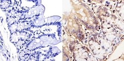
- Experimental details
- Immunohistochemistry-Paraffin: LAP1 Antibody (RL13) [NB120-2737] - Immunohistochemistry was performed on rat colon tissue. To expose target protein, antigen was retreived. Endogenous peroxidases were blocked in 3% H202-methanol for 15 minutes and tissues were blocked in 3% BSA-PBS for 30 minutes at room temperature. Cells were probed with a LAP1 mouse monoclonal antibody at a dilution of 1:20 overnight in a humidified chamber. Tissues were washed in PBST and detection was performed. DAB staining buffer was applied and tissues were counterstained with hematoxylin and prepped for mounting.

- Submitted by
- Novus Biologicals (provider)
- Main image

- Experimental details
- Immunohistochemistry: LAP1 Antibody (RL13) [NB120-2737] - Immunohistochemistry was performed on rat breast tissue. To expose target protein, antigen was retreived. Endogenous peroxidases were blocked in 3% H202-methanol for 15 minutes and tissues were blocked in 3% BSA-PBS for 30 minutes at room temperature. Cells were probed with a LAP1 mouse monoclonal antibody at a dilution of 1:50 overnight in a humidified chamber. Tissues were washed in PBST and detection was performed. DAB staining buffer was applied and tissues were counterstained with hematoxylin and prepped for mounting.
