 Explore
 Validate
 Learn
Learn Immunocytochemistry
Antibody data
- Antibody Data
- Antigen structure
- References [6]
- Comments [0]
- Validations
- Immunocytochemistry [1]
- Immunohistochemistry [3]
Submit
Validation data
Reference
Comment
Report error
- Product number
- MA1-074 - Provider product page

- Provider
- Invitrogen Antibodies
- Product name
- LAP1 Monoclonal Antibody (RL13)
- Antibody type
- Monoclonal
- Antigen
- Other
- Description
- MA1-074 detects lamin associated polypeptide (LAP) 1A, 1B & 1C from rat, human, and mouse samples. This antibody does not cross-react with LAP 2.
- Antibody clone number
- RL13
- Concentration
- 1 mg/mL
Submitted references SUN1 interacts with nuclear lamin A and cytoplasmic nesprins to provide a physical connection between the nuclear lamina and the cytoskeleton.
Lamin-binding fragment of LAP2 inhibits increase in nuclear volume during the cell cycle and progression into S phase.
Lamin-binding fragment of LAP2 inhibits increase in nuclear volume during the cell cycle and progression into S phase.
cDNA cloning and characterization of lamina-associated polypeptide 1C (LAP1C), an integral protein of the inner nuclear membrane.
Integral membrane proteins of the nuclear envelope interact with lamins and chromosomes, and binding is modulated by mitotic phosphorylation.
Integral membrane proteins specific to the inner nuclear membrane and associated with the nuclear lamina.
Haque F, Lloyd DJ, Smallwood DT, Dent CL, Shanahan CM, Fry AM, Trembath RC, Shackleton S
Molecular and cellular biology 2006 May;26(10):3738-51
Molecular and cellular biology 2006 May;26(10):3738-51
Lamin-binding fragment of LAP2 inhibits increase in nuclear volume during the cell cycle and progression into S phase.
Yang L, Guan T, Gerace L
The Journal of cell biology 1997 Dec 1;139(5):1077-87
The Journal of cell biology 1997 Dec 1;139(5):1077-87
Lamin-binding fragment of LAP2 inhibits increase in nuclear volume during the cell cycle and progression into S phase.
Yang L, Guan T, Gerace L
The Journal of cell biology 1997 Dec 1;139(5):1077-87
The Journal of cell biology 1997 Dec 1;139(5):1077-87
cDNA cloning and characterization of lamina-associated polypeptide 1C (LAP1C), an integral protein of the inner nuclear membrane.
Martin L, Crimaudo C, Gerace L
The Journal of biological chemistry 1995 Apr 14;270(15):8822-8
The Journal of biological chemistry 1995 Apr 14;270(15):8822-8
Integral membrane proteins of the nuclear envelope interact with lamins and chromosomes, and binding is modulated by mitotic phosphorylation.
Foisner R, Gerace L
Cell 1993 Jul 2;73(7):1267-79
Cell 1993 Jul 2;73(7):1267-79
Integral membrane proteins specific to the inner nuclear membrane and associated with the nuclear lamina.
Senior A, Gerace L
The Journal of cell biology 1988 Dec;107(6 Pt 1):2029-36
The Journal of cell biology 1988 Dec;107(6 Pt 1):2029-36
No comments: Submit comment
Supportive validation
- Submitted by
- Invitrogen Antibodies (provider)
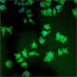
- Main image
- Experimental details
- Immunofluorescent analysis of LAP1 using anti-LAP1 monoclonal antibody (Product # MA1-074) shows staining in NS-1 Cells.

Supportive validation
- Submitted by
- Invitrogen Antibodies (provider)
- Main image

- Experimental details
- Immunohistochemistry was performed on rat breast tissue. To expose target protein, antigen was retreived using 10mM sodium citrate followed by microwave treatment for 8-15 minutes. Endogenous peroxidases were blocked in 3% H202-methanol for 15 minutes and tissues were blocked in 3% BSA-PBS for 30 minutes at room temperature. Cells were probed with a LAP1 mouse monoclonal antibody (Product # MA1-074) at a dilution of 1:50 overnight in a humidified chamber. Tissues were washed in PBST and detection was performed using a secondary antibody conjugated to HRP. DAB staining buffer was applied and tissues were counterstained with hematoxylin and prepped for mounting. Images were taken at 40X magnification.

- Submitted by
- Invitrogen Antibodies (provider)
- Main image

- Experimental details
- Immunohistochemistry was performed on rat colon tissue. To expose target protein, antigen was retreived using 10mM sodium citrate followed by microwave treatment for 8-15 minutes. Endogenous peroxidases were blocked in 3% H202-methanol for 15 minutes and tissues were blocked in 3% BSA-PBS for 30 minutes at room temperature. Cells were probed with a LAP1 mouse monoclonal antibody (Product # MA1-074) at a dilution of 1:20 overnight in a humidified chamber. Tissues were washed in PBST and detection was performed using a secondary antibody conjugated to HRP. DAB staining buffer was applied and tissues were counterstained with hematoxylin and prepped for mounting. Images were taken at 40X magnification.

- Submitted by
- Invitrogen Antibodies (provider)
- Main image

- Experimental details
- Immunohistochemistry was performed on rat lymph node tissue. To expose target protein, antigen was retreived using 10mM sodium citrate followed by microwave treatment for 8-15 minutes. Endogenous peroxidases were blocked in 3% H202-methanol for 15 minutes and tissues were blocked in 3% BSA-PBS for 30 minutes at room temperature. Cells were probed with a LAP1 mouse monoclonal antibody (Product # MA1-074) at a dilution of 1:50 overnight in a humidified chamber. Tissues were washed in PBST and detection was performed using a secondary antibody conjugated to HRP. DAB staining buffer was applied and tissues were counterstained with hematoxylin and prepped for mounting. Images were taken at 40X magnification.
