 Explore
 Validate
 Learn
Learn Western blot
Antibody data
- Antibody Data
- Antigen structure
- References [0]
- Comments [0]
- Validations
- Western blot [1]
- Immunohistochemistry [1]
- Flow cytometry [1]
Submit
Validation data
Reference
Comment
Report error
- Product number
- APC-125-25UL - Provider product page

- Provider
- Invitrogen Antibodies
- Product name
- Kir7.1 (extracellular) Polyclonal Antibody
- Antibody type
- Polyclonal
- Antigen
- Other
- Description
- Reconstitution: 1 X 25 µL double distilled water (DDW), depending on the sample size. The antibody ships as a lyophilized powder at room temperature. Upon arrival, it should be stored at -20C. The reconstituted solution can be stored at 4C for up to 1 week. For longer periods, small aliquots should be stored at -20C. Avoid multiple freezing and thawing. Centrifuge all antibody preparations before use (10000 x g 5 min).
- Reactivity
- Human, Mouse, Rat
- Host
- Rabbit
- Isotype
- IgG
- Vial size
- 25 µL
- Concentration
- 0.8 mg/mL
- Storage
- -20° C, Avoid Freeze/Thaw Cycles
No comments: Submit comment
Supportive validation
- Submitted by
- Invitrogen Antibodies (provider)
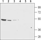
- Main image

- Experimental details
- Western blot analysis of rat brain membrane (lanes 1 and 4), mouse brain (lanes 2 and 5) and mouse kidney lysates (lanes 3 and 6): - 1-3. Anti-Kir7.1 (extracellular) Antibody (#APC-125), (1:200).4-6. Anti-Kir7.1 (extracellular) Antibody , preincubated with Kir7.1 (extracellular) Blocking Peptide (#BLP-PC125).
Supportive validation
- Submitted by
- Invitrogen Antibodies (provider)
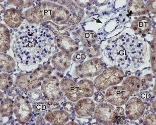
- Main image

- Experimental details
- Expression of Kir7.1 in rat kidney - Immunohistochemical staining of paraffin embedded section of rat kidney using Anti-Kir7.1 (extracellular) Antibody (#APC-125), (1:100). Staining is present in both distal (DT) and proximal (PT) tubules and in the collecting ducts (CD) in the renal cortex. Note that staining is absent both inglomeruli (Gl) and blood vessels (arrow). Hematoxilin is used as the counterstain.
Supportive validation
- Submitted by
- Invitrogen Antibodies (provider)
- Main image

- Experimental details
- Cell surface detection of Kir7.1 in live intact Jurkat (human T cell leukemia) cell line: - (black) Cells + Goat- Anti-Rabbit-FITC. (green) Cells + Anti-Kir7.1 (extracellular) Antibody (#APC-125), (1:20) + Goat- Anti-Rabbit-FITC.
