Explore
 Validate
 Learn
LearnPA3-118
antibody from Invitrogen Antibodies
Targeting: PRG4
bG174L6.2, CACP, FLJ32635, HAPO, JCAP, MSF, SZP
 Western blot
Antibody data
- Antibody Data
- Antigen structure
- References [7]
- Comments [0]
- Validations
- Western blot [1]
- Other assay [5]
Submit
Validation data
Reference
Comment
Report error
- Product number
- PA3-118 - Provider product page

- Provider
- Invitrogen Antibodies
- Product name
- Lubricin Polyclonal Antibody
- Antibody type
- Polyclonal
- Antigen
- Synthetic peptide
- Description
- PA3-118 detects Lubricin in human samples. PA3-118 has been successfully used in Western blot procedures. By Western blot, this antibody detects a 280 kDa protein representing recombinant human Lubricin. PA3-118 works best on purified or recombinant samples under non-reducing conditions. PA3-118 immunogen is a synthetic peptide corresponding to residues C L(1356) P N I R K Q P D G Y D Y Y A F S K D Q(1374) of human Lubricin. This polyclonal antibody has been referred to as: JSCLPN.
- Reactivity
- Human
- Host
- Rabbit
- Isotype
- IgG
- Vial size
- 100 µg
- Concentration
- 1 mg/mL
- Storage
- -20° C, Avoid Freeze/Thaw Cycles
Submitted references Proteoglycan 4 (PRG4) expression and function in dry eye associated inflammation.
Synovial fluid lubricin and hyaluronan are altered in equine osteochondral fragmentation, cartilage impact injury, and full-thickness cartilage defect models.
Prg4 prevents osteoarthritis induced by dominant-negative interference of TGF-ß signaling in mice.
Temporal changes in synovial fluid composition and elastoviscous lubrication in the equine carpal fracture model.
Lubricin binds cartilage proteins, cartilage oligomeric matrix protein, fibronectin and collagen II at the cartilage surface.
Hox11 genes are required for regional patterning and integration of muscle, tendon and bone.
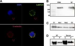
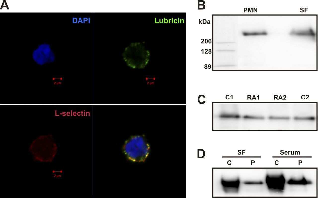
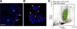
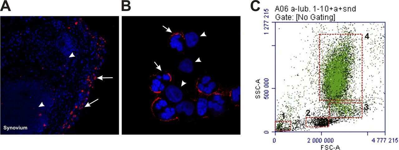
Human synovial lubricin expresses sialyl Lewis x determinant and has L-selectin ligand activity.
Menon NG, Goyal R, Lema C, Woods PS, Tanguay AP, Morin AA, Das N, Jay GD, Krawetz RJ, Dufour A, Shapiro LH, Redfern RL, Ghosh M, Schmidt TA
Experimental eye research 2021 Jul;208:108628
Experimental eye research 2021 Jul;208:108628
Synovial fluid lubricin and hyaluronan are altered in equine osteochondral fragmentation, cartilage impact injury, and full-thickness cartilage defect models.
Peal BT, Gagliardi R, Su J, Fortier LA, Delco ML, Nixon AJ, Reesink HL
Journal of orthopaedic research : official publication of the Orthopaedic Research Society 2020 Aug;38(8):1826-1835
Journal of orthopaedic research : official publication of the Orthopaedic Research Society 2020 Aug;38(8):1826-1835
Prg4 prevents osteoarthritis induced by dominant-negative interference of TGF-ß signaling in mice.
Chavez RD, Sohn P, Serra R
PloS one 2019;14(1):e0210601
PloS one 2019;14(1):e0210601
Temporal changes in synovial fluid composition and elastoviscous lubrication in the equine carpal fracture model.
Feeney E, Peal BT, Inglis JE, Su J, Nixon AJ, Bonassar LJ, Reesink HL
Journal of orthopaedic research : official publication of the Orthopaedic Research Society 2019 May;37(5):1071-1079
Journal of orthopaedic research : official publication of the Orthopaedic Research Society 2019 May;37(5):1071-1079
Lubricin binds cartilage proteins, cartilage oligomeric matrix protein, fibronectin and collagen II at the cartilage surface.
Flowers SA, Zieba A, Örnros J, Jin C, Rolfson O, Björkman LI, Eisler T, Kalamajski S, Kamali-Moghaddam M, Karlsson NG
Scientific reports 2017 Oct 13;7(1):13149
Scientific reports 2017 Oct 13;7(1):13149
Hox11 genes are required for regional patterning and integration of muscle, tendon and bone.
Swinehart IT, Schlientz AJ, Quintanilla CA, Mortlock DP, Wellik DM
Development (Cambridge, England) 2013 Nov;140(22):4574-82
Development (Cambridge, England) 2013 Nov;140(22):4574-82
Human synovial lubricin expresses sialyl Lewis x determinant and has L-selectin ligand activity.
Jin C, Ekwall AK, Bylund J, Björkman L, Estrella RP, Whitelock JM, Eisler T, Bokarewa M, Karlsson NG
The Journal of biological chemistry 2012 Oct 19;287(43):35922-33
The Journal of biological chemistry 2012 Oct 19;287(43):35922-33
No comments: Submit comment
Supportive validation
- Submitted by
- Invitrogen Antibodies (provider)
- Main image

- Experimental details
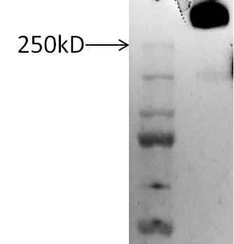
- Western blot analysis of Lubricin was performed by loading 3 µg of non-reduced human synovial fluid per well onto a SDS-PAGE gel. Proteins were transferred to a membrane. The membrane was probed with a Lubricin polyclonal antibody (Product # PA3-118) at a concentration of 2 µg/mL diluted in the antibody diluent provided in the Fast Western Blot Kit (Product # 35050), followed by the optimized HRP reagent from the same kit (diluted by adding 150 µL of HRP reagent to 10 mL of reagent diluent). Chemiluminescent detection was performed using SuperSignal West Dura (Product # 34075).
Supportive validation
- Submitted by
- Invitrogen Antibodies (provider)
- Main image
- Experimental details
- NULL

- Submitted by
- Invitrogen Antibodies (provider)
- Main image
- Experimental details
- NULL
- Submitted by
- Invitrogen Antibodies (provider)
- Main image
- Experimental details
- NULL
- Submitted by
- Invitrogen Antibodies (provider)
- Main image
- Experimental details
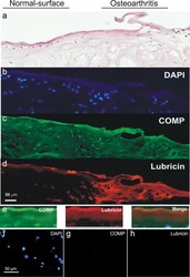
- Figure 1 Immunohistochemical co-localisation of lubricin and COMP. Dual antibody immunofluorescence on OA cartilage biopsy cryosections with anti-lubricin (P3-118) and anti-COMP (mAb 16F12) antibodies. Section shows an area of undisrupted cartilage surface (left side) and an area more severely affected by OA including a tear (right side). ( a ) Haemotoxylin and esosin staining. ( b ) DAPI staining for nuclei of chondrocytes. ( c ) COMP (green) was constantly distributed over the section with greater intensity on the superficial zone. ( d ) Lubricin (red) was present on the superficial zone of the cartilage and into the superficial zone in the area of the tissue with OA degradation at the surface. ( e ) Cartilage surface alone including COMP antibody staining, lubricin antibody staining and merged image showing co-localisation of COMP and lubricin on cartilage surface. Negative controls of dual antibody immunofluorescence analysis. The specificity of the staining was verified using matched isotype negative controls or control serum at the same concentration as the primary antibodies. ( f ) Negative control showing DAPI staining. ( g ) Negative control for the anti-COMP antibody. ( h ) Negative control for the anti-lubricin antibody.

- Submitted by
- Invitrogen Antibodies (provider)
- Main image
- Experimental details
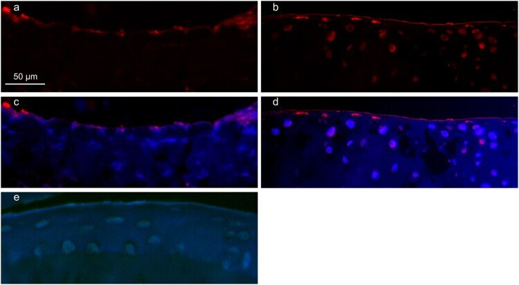
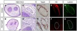
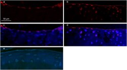
- Figure 2 Immunohistochemical localisation of lubricin in WT and COMP KO mice paw joint cartilage tissue. ( a ) WT mouse tissue stained with rabbit anti-lubricin (P3-118) visualised with Rhodamine Red-X shows a diffuse lubricin layer at the cartilage surface. ( b ) COMP KO mouse tissue stained with rabbit anti-lubricin (P3-118) visualised with Rhodamine Red-X showing a discrete, distinct layer of lubricin at the cartilage surface. ( c ) WT mouse tissue stained with rabbit anti-lubricin (P3-118) visualised with Rhodamine Red-X also showing DAPI staining. ( d ) COMP KO mouse tissue stained with rabbit anti-lubricin (P3-118) visualised with Rhodamine Red-X also showing DAPI staining. ( e ) Negative control of WT mouse paw tissue performed by omitting primary antibody.