Antibody data
- Antibody Data
- Antigen structure
- References [1]
- Comments [0]
- Validations
- Western blot [1]
- Immunohistochemistry [1]
Submit
Validation data
Reference
Comment
Report error
- Product number
- NB600-472 - Provider product page

- Provider
- Novus Biologicals
- Proper citation
- Novus Cat#NB600-472, RRID:AB_10001053
- Product name
- Rabbit Polyclonal Ubiquitin-activating Enzyme/UBE1 Antibody
- Antibody type
- Polyclonal
- Description
- IgG purified. UBE1
- Reactivity
- Human
- Host
- Rabbit
- Isotype
- IgG
- Vial size
- 0.25 mg
- Storage
- Store at 4C short term. Aliquot and store at -20C long term. Avoid freeze-thaw cycles.
Submitted references A cohesin-RAD21 interactome.
Panigrahi AK, Zhang N, Otta SK, Pati D
The Biochemical journal 2012 Mar 15;442(3):661-70
The Biochemical journal 2012 Mar 15;442(3):661-70
No comments: Submit comment
Supportive validation
- Submitted by
- Novus Biologicals (provider)
- Main image

- Experimental details
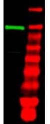
- Western Blot: Ubiquitin-activating Enzyme/UBE1 Antibody [NB600-472] - Shows detection of a band at ~118 kDa corresponding to UBE1 (lane 1 800 nm channel). Approximately 35 ug of an A431 whole cell lysate was separated on a 4-20% Tris-Glycine gel by SDS-PAGE and transferred onto nitrocellulose. After blocking the membrane was probed with the primary antibody diluted to 1:1,000. Incubation was for 2 h at room temperature followed by washes and reaction with a 1:10,000 dilution of IRDye (TM) 800 conjugated Gt-a-Rabbit IgG [H&L] MX10 for 45 min at room temperature. Molecular weight markers are shown in lane 2 (700 nm channel).
Supportive validation
- Submitted by
- Novus Biologicals (provider)
- Main image

- Experimental details
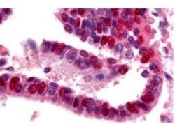
- Immunohistochemistry: Ubiquitin-activating Enzyme/UBE1 Antibody [NB600-472] - Used at a 10 ug/ml to detect UBE1 in a variety of tissues including adrenal, breast, colon (epithelium), kidney, liver, lung (respiratory epithelium), ovary (oocyte and endothelium), pancreas (islet and exocrine), placenta, prostate (epithelium), skin (epithelium), spleen (lymphocytes), stomach (chief), testis, thymus, tonsil, and uterus (glandular, stroma). In many cells a punctate nuclear staining was observed. Other cells showed both cytoplasmic and nuclear staining. This image shows UBE1 staining of human lung tissue. Tissue was formalin-fixed and paraffin embedded. Personal Communication, Tina Roush, LifeSpanBiosciences, Seattle, WA.
 Explore
 Validate
 Learn
Learn Western blot
 ELISA
ELISA