Explore
 Validate
 Learn
Learn Western blot
 ELISA
ELISAAntibody data
- Antibody Data
- Antigen structure
- References [5]
- Comments [0]
- Validations
- Western blot [12]
- ELISA [1]
- Immunocytochemistry [2]
- Immunoprecipitation [1]
- Immunohistochemistry [1]
- Flow cytometry [1]
Submit
Validation data
Reference
Comment
Report error
- Product number
- GTX104557 - Provider product page

- Provider
- GeneTex
- Proper citation
- GeneTex Cat#GTX104557, RRID:AB_1950436
- Product name
- Glypican 1 antibody [N3C3]
- Antibody type
- Polyclonal
- Reactivity
- Human, Mouse, Rat
- Host
- Rabbit
Submitted references Abnormal ER quality control of neural GPI-anchored proteins via dysfunction in ER export processing in the frontal cortex of elderly subjects with schizophrenia.
Sphingosine-1-phosphate improves endothelialization with reduction of thrombosis in recellularized human umbilical vein graft by inhibiting syndecan-1 shedding in vitro.
Elevated glypican-1 expression is associated with an unfavorable prognosis in pancreatic ductal adenocarcinoma.
α3 Chains of type V collagen regulate breast tumour growth via glypican-1.
Differential expression of glypican-1 in ameloblastoma variants.
Kim P, Scott MR, Meador-Woodruff JH
Translational psychiatry 2019 Jan 16;9(1):6
Translational psychiatry 2019 Jan 16;9(1):6
Sphingosine-1-phosphate improves endothelialization with reduction of thrombosis in recellularized human umbilical vein graft by inhibiting syndecan-1 shedding in vitro.
Hsia K, Yang MJ, Chen WM, Yao CL, Lin CH, Loong CC, Huang YL, Lin YT, Lander AD, Lee H, Lu JH
Acta biomaterialia 2017 Mar 15;51:341-350
Acta biomaterialia 2017 Mar 15;51:341-350
Elevated glypican-1 expression is associated with an unfavorable prognosis in pancreatic ductal adenocarcinoma.
Lu H, Niu F, Liu F, Gao J, Sun Y, Zhao X
Cancer medicine 2017 Jun;6(6):1181-1191
Cancer medicine 2017 Jun;6(6):1181-1191
α3 Chains of type V collagen regulate breast tumour growth via glypican-1.
Huang G, Ge G, Izzi V, Greenspan DS
Nature communications 2017 Jan 19;8:14351
Nature communications 2017 Jan 19;8:14351
Differential expression of glypican-1 in ameloblastoma variants.
Bologna-Molina R, Mosqueda-Taylor A, Molina-Frechero N
Applied immunohistochemistry & molecular morphology : AIMM 2015 Feb;23(2):153-60
Applied immunohistochemistry & molecular morphology : AIMM 2015 Feb;23(2):153-60
No comments: Submit comment
Enhanced validation
Supportive validation
- Submitted by
- GeneTex (provider)
- Enhanced method
- Genetic validation
- Main image
- Experimental details
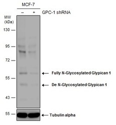
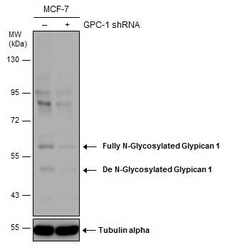
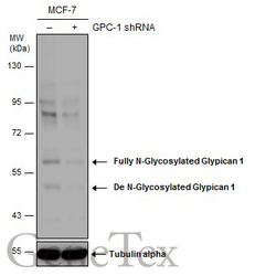
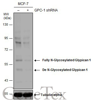
- Non-transfected (¡V) and transfected (+) MCF-7 whole cell extracts (50 ?g) were separated by 7.5% SDS-PAGE, and the membrane was blotted with Glypican 1 antibody [N3C3] (GTX104557) diluted at 1:1500. The HRP-conjugated anti-rabbit IgG antibody (GTX213110-01) was used to detect the primary antibody.
Supportive validation
- Submitted by
- GeneTex (provider)
- Main image

- Experimental details
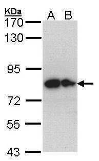
- Sample (30 ug of whole cell lysate) A: Hep G2 (GTX27900) B: Molt-4 (GTX27912) 7.5% SDS PAGE GTX104557 diluted at 1:1000
- Validation comment
- WB
- Submitted by
- GeneTex (provider)
- Main image
- Experimental details
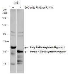
- Untreated (¡V) and treated (+) A431 whole cell extracts (30 ?g) were separated by 7.5% SDS-PAGE, and the membrane was blotted with Glypican 1 antibody [N3C3] (GTX104557) diluted at 1:500. The HRP-conjugated anti-rabbit IgG antibody (GTX213110-01) was used to detect the primary antibody.
- Submitted by
- GeneTex (provider)
- Main image
- Experimental details
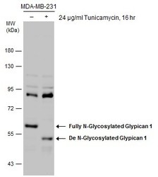
- Untreated (¡V) and treated (+) MDA-MB-231 whole cell extracts (30 ?g) were separated by 7.5% SDS-PAGE, and the membrane was blotted with Glypican 1 antibody [N3C3] (GTX104557) diluted at 1:1500. The HRP-conjugated anti-rabbit IgG antibody (GTX213110-01) was used to detect the primary antibody.
- Submitted by
- GeneTex (provider)
- Main image
- Experimental details
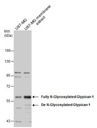
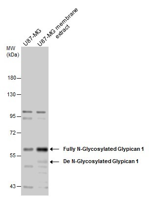
- U87-MG whole cell and membrane extracts (30 ?g) were separated by 7.5% SDS-PAGE, and the membrane was blotted with Glypican 1 antibody [N3C3] (GTX104557) diluted at 1:1500. The HRP-conjugated anti-rabbit IgG antibody (GTX213110-01) was used to detect the primary antibody.
- Submitted by
- GeneTex (provider)
- Main image
- Experimental details
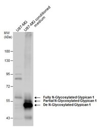
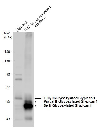
- U87-MG whole cell extract and conditioned medium (30 ?g) were separated by 7.5% SDS-PAGE, and the membrane was blotted with Glypican 1 antibody [N3C3] (GTX104557) diluted at 1:1500. The HRP-conjugated anti-rabbit IgG antibody (GTX213110-01) was used to detect the primary antibody.
- Submitted by
- GeneTex (provider)
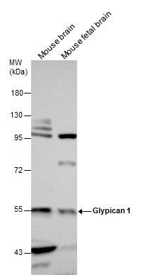
- Main image
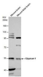
- Experimental details
- Mouse tissue extracts (50 ?g) were separated by 7.5% SDS-PAGE, and the membrane was blotted with Glypican 1 antibody [N3C3] (GTX104557) diluted at 1:1500. The HRP-conjugated anti-rabbit IgG antibody (GTX213110-01) was used to detect the primary antibody.
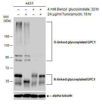
- Submitted by
- GeneTex (provider)
- Main image
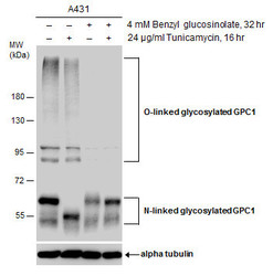
- Experimental details
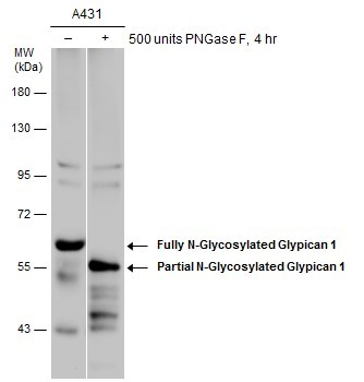
- Untreated (¡V) and treated (+) A431 whole cell extracts (30 ?g) were separated by 7.5% SDS-PAGE, and the membrane was blotted with Glypican 1 antibody [N3C3] (GTX104557) diluted at 1:3000. The HRP-conjugated anti-rabbit IgG antibody (GTX213110-01) was used to detect the primary antibody.
- Submitted by
- GeneTex (provider)
- Main image
- Experimental details
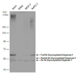
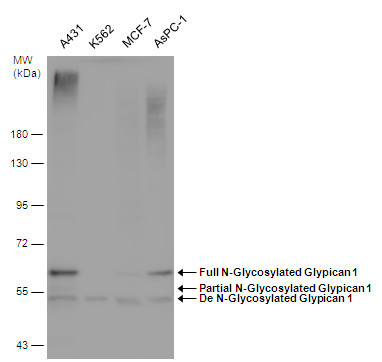
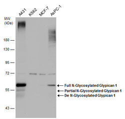
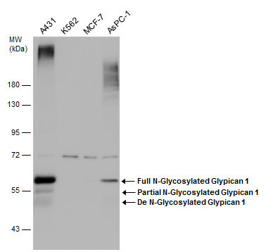
- Various whole cell extracts (30 ?g) were separated by 7.5% SDS-PAGE, and the membrane was blotted with Glypican 1 antibody [N3C3] (GTX104557) diluted at 1:1500. The HRP-conjugated anti-rabbit IgG antibody (GTX213110-01) was used to detect the primary antibody.
- Submitted by
- GeneTex (provider)
- Main image
- Experimental details
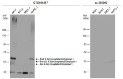
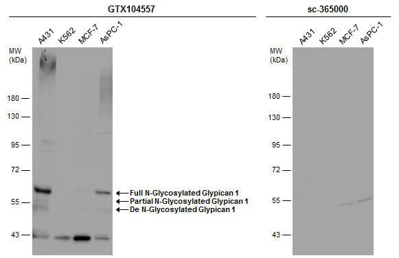
- Various whole cell extracts (30 ?g) were separated by 7.5% SDS-PAGE, and the membranes were blotted with Glypican 1 antibody [N3C3] (GTX104557) diluted at 1:500 and competitor's antibody (sc-365000) diluted at 1:500. The HRP-conjugated anti-rabbit IgG antibody (GTX213110-01) was used to detect the primary antibody.
- Submitted by
- GeneTex (provider)
- Main image
- Experimental details
- Non-transfected (¡V) and transfected (+) MCF-7 whole cell extracts (50 ?g) were separated by 7.5% SDS-PAGE, and the membrane was blotted with Glypican 1 antibody [N3C3] (GTX104557) diluted at 1:1500. The HRP-conjugated anti-rabbit IgG antibody (GTX213110-01) was used to detect the primary antibody.
- Submitted by
- GeneTex (provider)
- Main image
- Experimental details
- Various whole cell extracts (30 ?g) were separated by 7.5% SDS-PAGE, and the membrane was blotted with Glypican 1 antibody [N3C3] (GTX104557) diluted at 1:1500.
Supportive validation
- Submitted by
- GeneTex (provider)
- Main image
- Experimental details
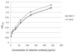
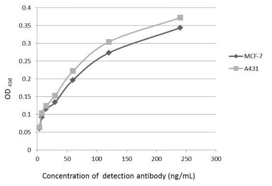
- An ELISA plate is coated with MCF-7 and A431 cells. The coated cells are detected with Glypican 1 antibody (GTX104557) at concentration ranged from 7.5 to 240 ng/mL.
Supportive validation
- Submitted by
- GeneTex (provider)
- Main image

- Experimental details
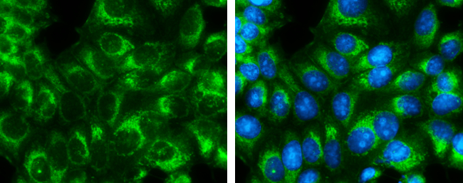
- Glypican 1 antibody [N3C3] detects Glypican 1 protein at endoplasmic reticulum by immunofluorescent analysis.Sample: MCF7 cells were fixed in ice-cold MeOH for 5 min.Green: Glypican 1 protein stained by Glypican 1 antibody [N3C3] (GTX104557) diluted at 1:400.Blue: Hoechst 33342 staining.
- Submitted by
- GeneTex (provider)
- Main image

- Experimental details
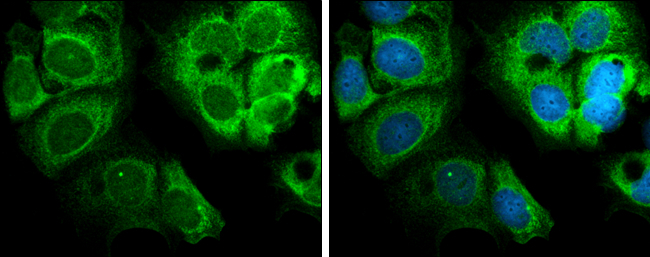
- Glypican 1 antibody [N3C3] detects Glypican 1 protein at cytoplasm by immunofluorescent analysis.Sample: MCF7 cells were fixed in ice-cold MeOH for 5 min.Green: Glypican 1 protein stained by Glypican 1 antibody [N3C3] (GTX104557) diluted at 1:500.Blue: Hoechst 33342 staining.
Supportive validation
- Submitted by
- GeneTex (provider)
- Main image
- Experimental details
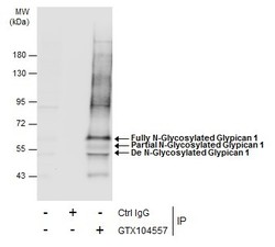
- Immunoprecipitation of Glypican 1 protein from MCF-7 whole cell extracts using 5 £gg of Glypican 1 antibody [N3C3] (GTX104557).Western blot analysis was performed using Glypican 1 antibody [N3C3] (GTX104557).EasyBlot anti-Rabbit IgG (GTX221666-01) was used as a secondary reagent.
Supportive validation
- Submitted by
- GeneTex (provider)
- Main image
- Experimental details
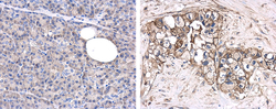
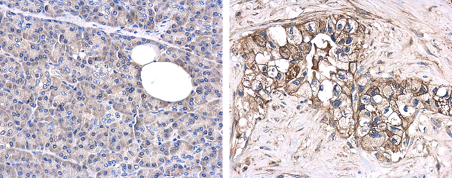
- Immunohistochemical microscopy analysis of paraffin-embedded human normal pancreas (left) or pancreatic adenocarcinoma (grade II) (right) tissues using Glypican-1 antibody [N3C3] (GTX104557) at a 1:250 dilution.
Supportive validation
- Submitted by
- GeneTex (provider)
- Main image
- Experimental details
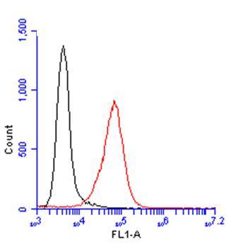
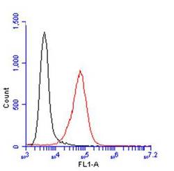
- Glypican 1 antibody [N3C3] (GTX104557) detects Glypican 1 protein by flow cytometry analysis. Sample: A431 cell. Black: Unlabelled sample was used as a control. Red: Glypican 1 antibody [N3C3] (GTX104557) dilution: 1:100. Acquisition of 20,000 events were collected using a Dylight 488-conjugated secondary antibody for FACS analysis.