 Explore
 Validate
 Learn
Learn Western blot
Antibody data
- Antibody Data
- Antigen structure
- References [1]
- Comments [0]
- Validations
- Western blot [1]
- Immunocytochemistry [3]
- Immunohistochemistry [2]
- Flow cytometry [1]
- Other assay [1]
Submit
Validation data
Reference
Comment
Report error
- Product number
- PA5-86043 - Provider product page

- Provider
- Invitrogen Antibodies
- Product name
- Glypican 1 Polyclonal Antibody
- Antibody type
- Polyclonal
- Antigen
- Synthetic peptide
- Reactivity
- Human
- Host
- Rabbit
- Isotype
- IgG
- Vial size
- 100 µL
- Concentration
- 1 mg/mL
- Storage
- Store at 4°C short term. For long term storage, store at -20°C, avoiding freeze/thaw cycles.
Submitted references The proteomic analysis of breast cell line exosomes reveals disease patterns and potential biomarkers.
Risha Y, Minic Z, Ghobadloo SM, Berezovski MV
Scientific reports 2020 Aug 11;10(1):13572
Scientific reports 2020 Aug 11;10(1):13572
No comments: Submit comment
Supportive validation
- Submitted by
- Invitrogen Antibodies (provider)
- Main image

- Experimental details
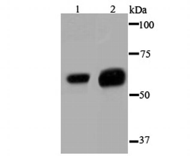
- Western blot analysis of Glypican 1 in different lysates using a Polyclonal antibody (Product #PA5-86043) at a dilution of 1:1,000. Positive control: Lane1: Hela, Lane 2: SK-Br-3.
Supportive validation
- Submitted by
- Invitrogen Antibodies (provider)
- Main image

- Experimental details
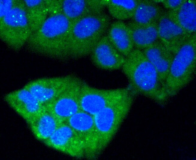
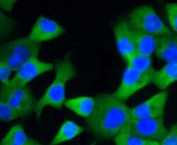
- Immunocytochemical analysis of Glypican 1 in Hela cells using a Glypican 1 Polyclonal antibody (Product # PA5-86043) as seen in green. The nuclear counter stain is DAPI (blue). Cells were fixed in paraformaldehyde, permeabilised with 0.25% Triton X100/PBS.
- Submitted by
- Invitrogen Antibodies (provider)
- Main image
- Experimental details
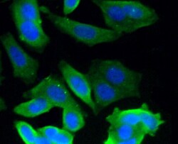
- Immunocytochemical analysis of Glypican 1 in MCF-7 cells using a Glypican 1 Polyclonal antibody (Product # PA5-86043) as seen in green. The nuclear counter stain is DAPI (blue). Cells were fixed in paraformaldehyde, permeabilised with 0.25% Triton X100/PBS.
- Submitted by
- Invitrogen Antibodies (provider)
- Main image
- Experimental details
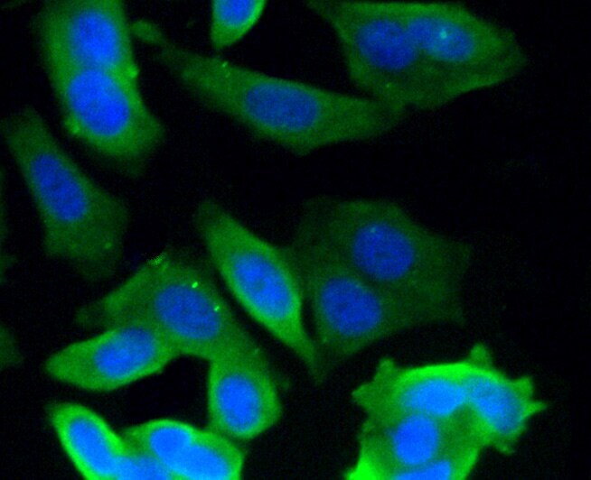
- Immunocytochemical analysis of Glypican 1 in PANC-1 cells using a Glypican 1 Polyclonal antibody (Product # PA5-86043) as seen in green. The nuclear counter stain is DAPI (blue). Cells were fixed in paraformaldehyde, permeabilised with 0.25% Triton X100/PBS.

Supportive validation
- Submitted by
- Invitrogen Antibodies (provider)
- Main image

- Experimental details
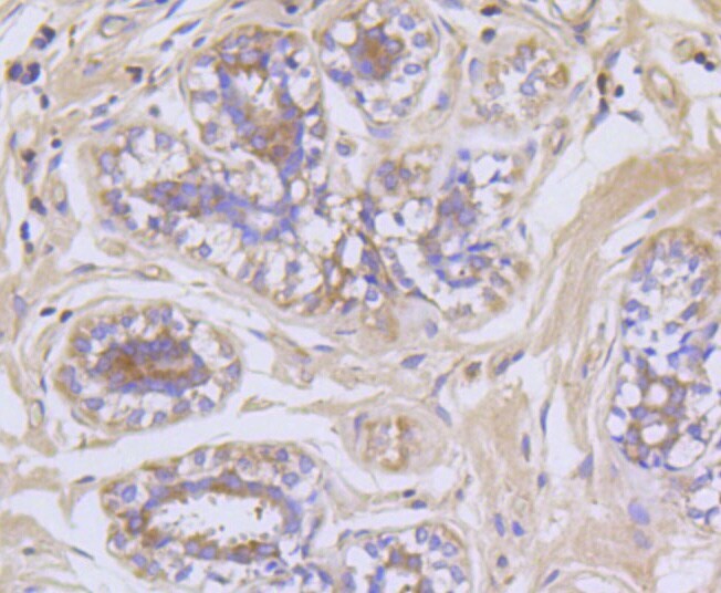
- Immunohistochemical analysis of Glypican 1 of paraffin-embedded Human breast tissue using a Glypican-1 Polyclonal antibody (Product #PA5-86043). Counter stained with hematoxylin.
- Submitted by
- Invitrogen Antibodies (provider)
- Main image

- Experimental details
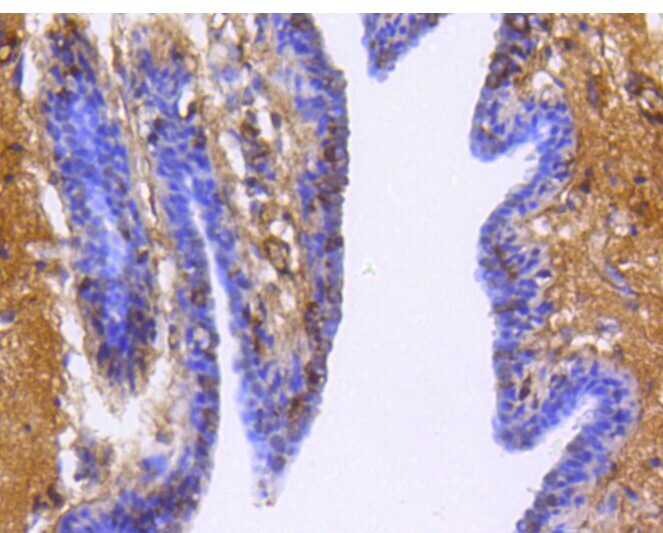
- Immunohistochemical analysis of Glypican 1 of paraffin-embedded Human breast cancer tissue using a Glypican-1 Polyclonal antibody (Product #PA5-86043). Counter stained with hematoxylin.
Supportive validation
- Submitted by
- Invitrogen Antibodies (provider)
- Main image
- Experimental details
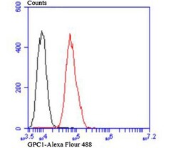
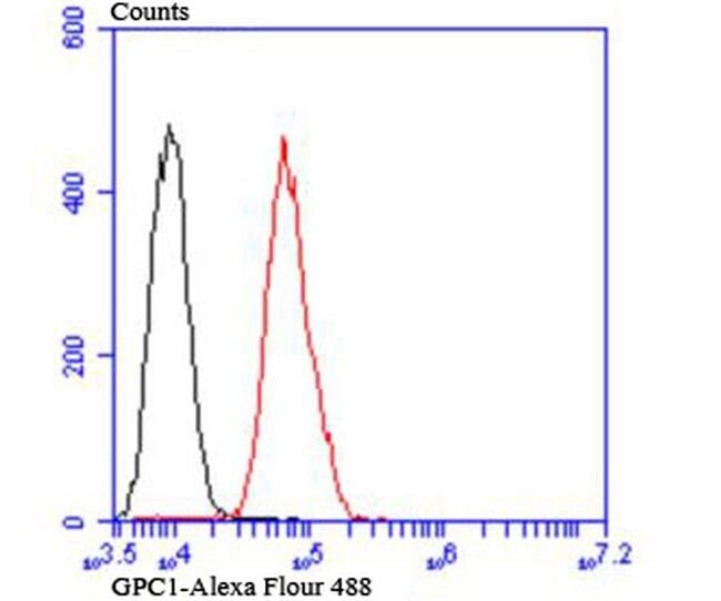
- Flow Cytometric analysis of Glypican 1 in MCF-7 cells using a Glypican 1 Polyclonal Antibody (Product # PA5-86043) at a dilution of 1:100, as seen in red compared with an unlabelled control (cells without incubation with primary antibody; black).
Supportive validation
- Submitted by
- Invitrogen Antibodies (provider)
- Main image
- Experimental details
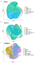
- Figure 5 tSNE plots of exosomes analyzed by FACS showing expression levels of ( a ) GPC-1 ( b ) GLUT-1 and ( c ) ADAM10 along with CD81 and CD63 on the surface of BC-derived exosomes.
