Explore
 Validate
 Learn
Learn Western blot
Antibody data
- Antibody Data
- Antigen structure
- References [1]
- Comments [0]
- Validations
- Western blot [14]
- ELISA [2]
- Immunocytochemistry [1]
- Immunohistochemistry [2]
- Flow cytometry [1]
- Other assay [2]
Submit
Validation data
Reference
Comment
Report error
- Product number
- PA5-28055 - Provider product page

- Provider
- Invitrogen Antibodies
- Product name
- Glypican 1 Polyclonal Antibody
- Antibody type
- Polyclonal
- Antigen
- Recombinant full-length protein
- Description
- Recommended positive controls: A431, K562, MCF-7, AsPC-1, MDA-MB-231, MDA-MB-231 (24 µg/mL Tunicamycin for 16hr), U87-MG, U87-MG membrane extracts, U87-MG conditioned medium, Mouse brain, mouse fetal brain, A431 (500 units PNGase F for 4 hr), A431 (24 µg/mL Tunicamycin treatment for 16hr), A431 (4 mM Benzyl glucosinolate treatment for 32hr), A431(24 µg/mL Tunicamycin treatment for 16hr and 4 mM Benzyl glucosinolate treatment for 32hr).
- Concentration
- 0.58 mg/mL
Submitted references Characterization of nucleic acids from extracellular vesicle-enriched human sweat.
Bart G, Fischer D, Samoylenko A, Zhyvolozhnyi A, Stehantsev P, Miinalainen I, Kaakinen M, Nurmi T, Singh P, Kosamo S, Rannaste L, Viitala S, Hiltunen J, Vainio SJ
BMC genomics 2021 Jun 9;22(1):425
BMC genomics 2021 Jun 9;22(1):425
No comments: Submit comment
Supportive validation
- Submitted by
- Invitrogen Antibodies (provider)
- Main image
- Experimental details
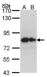
- Western Blot analysis using anti-Glypican 1 Polyclonal Antibody on the following cells: (30 µg of whole cell lysate) A: Hep G2 cell lysate B: Molt-4 cell lysate. Primary antibody (Product # PA5-28055) diluted at 1:1000

- Submitted by
- Invitrogen Antibodies (provider)
- Main image
- Experimental details
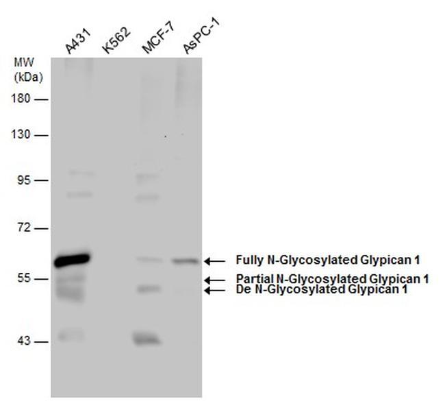
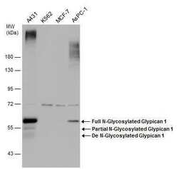
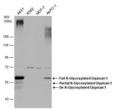
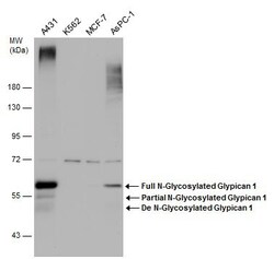
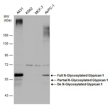
- Western Blot analysis of Glypican 1 was performed by loading 30 µg of (Left - Right) A431, K562, MCF-7, AsPC1 whole cell extracts per well into a 7.5% SDS-PAGE gel. After protein transfer, the membrane was probed with a polyclonal Gypican-1 antibody (N3C3) (Product # PA5-28055) at a dilution of 1:1500.
- Submitted by
- Invitrogen Antibodies (provider)
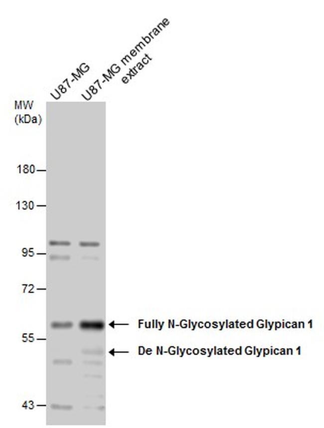
- Main image
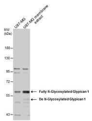
- Experimental details
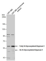
- Western Blot analysis of Glypican 1 was performed by loading 30 µg of (Left - Right) U87-MG whole cell and U87-MG membrane extracts per well into a 7.5% SDS-PAGE gel. After protein transfer, the membrane was probed with a polyclonal Gypican-1 antibody (N3C3) (Product # PA5-28055) at a dilution of 1:1500.
- Submitted by
- Invitrogen Antibodies (provider)
- Main image
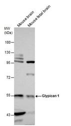
- Experimental details
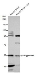
- Western Blot analysis of Glypican 1 was performed by loading 50 µg of (Left - Right) Mouse brane and Mouse fetal brain extracts per well into a 7.5% SDS-PAGE gel. After protein transfer, the membrane was probed with a polyclonal Gypican-1 antibody (N3C3) (Product # PA5-28055) at a dilution of 1:1500.

- Submitted by
- Invitrogen Antibodies (provider)
- Main image
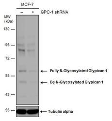
- Experimental details
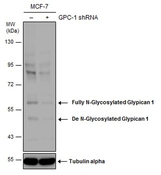
- Western Blot analysis of Glypican 1 was performed by loading 50 µg of (Left)Non-transfected MCF-7 whole cell extracts and (Right) MCF-7 whole cell extracts transfected with GPC-1 shRNA into a 7.5% SDS-PAGE gel. After protein transfer, the membrane was probed with a polyclonal Gypican-1 antibody (N3C3) (Product # PA5-28055) at a dilution of 1:1500.
- Submitted by
- Invitrogen Antibodies (provider)
- Main image
- Experimental details
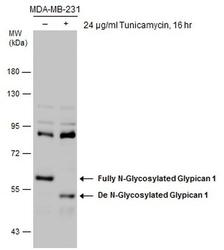
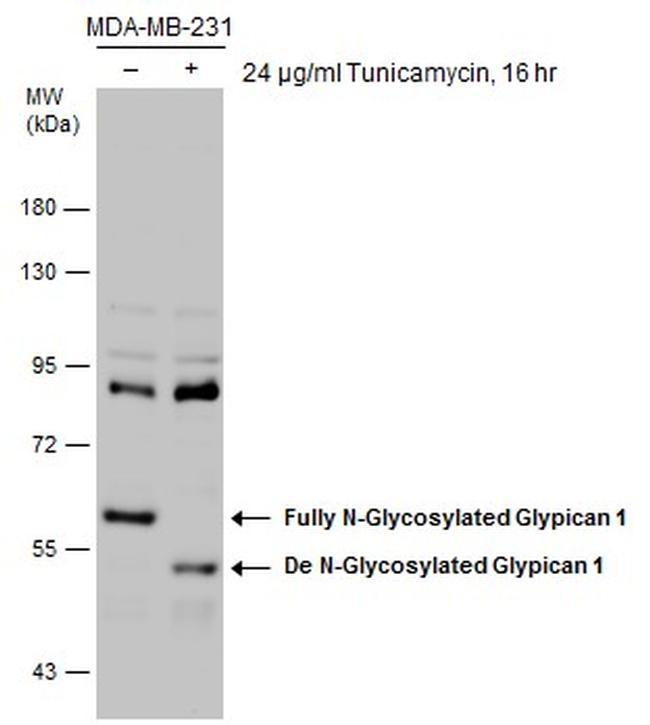
- Western Blot analysis of Glypican 1 was performed by loading 30 µg of (Left)untreated MDA-MB-231 whole cell extracts and (Right) MDA-MB-231 whole cell extracts treated with 24 µg/mL Tynicamycin for 16 hours into a 7.5% SDS-PAGE gel. After protein transfer, the membrane was probed with a polyclonal Gypican-1 antibody (N3C3) (Product # PA5-28055) at a dilution of 1:1500.
- Submitted by
- Invitrogen Antibodies (provider)
- Main image
- Experimental details
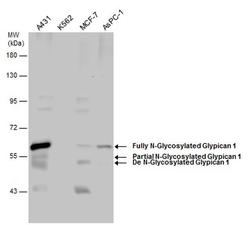
- Western Blot analysis of Glypican 1 was performed by separating 30 µg of various whole cell extracts by 7.5% SDS-PAGE. Proteins were transferred to a membrane and probed with a Glypican 1 Polyclonal Antibody (Product # PA5-28055) at a dilution of 1:1500.
- Submitted by
- Invitrogen Antibodies (provider)
- Main image
- Experimental details
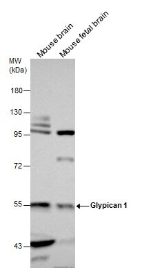
- Western Blot analysis of Glypican 1 was performed by separating 50 µg of Mouse tissue extracts by 7.5% SDS-PAGE. Proteins were transferred to a membrane and probed with a Glypican 1 Polyclonal Antibody (Product # PA5-28055) at a dilution of 1:1500. The HRP-conjugated anti-rabbit IgG antibody was used to detect the primary antibody.
- Submitted by
- Invitrogen Antibodies (provider)
- Main image
- Experimental details
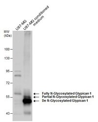
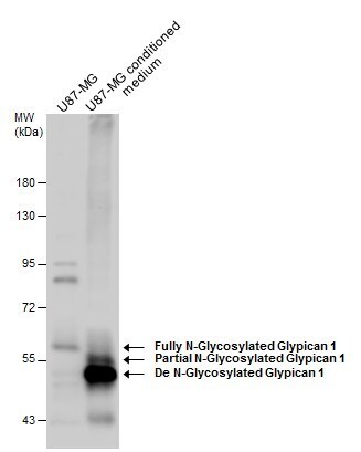
- Western Blot analysis of Glypican 1 was performed by separating 30 µg of U87-MG whole cell extracts and conditioned medium by 7.5% SDS-PAGE. Proteins were transferred to a membrane and probed with a Glypican 1 Polyclonal Antibody (Product # PA5-28055) at a dilution of 1:1500. The HRP-conjugated anti-rabbit IgG antibody was used to detect the primary antibody.
- Submitted by
- Invitrogen Antibodies (provider)
- Main image
- Experimental details
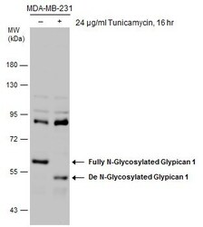
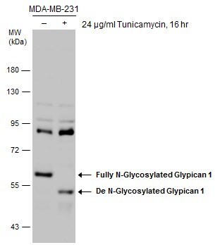
- Western Blot analysis of Glypican 1 was performed by separating 30 µg of untreated (–) and treated (+) MDA-MB-231 whole cell extracts by 7.5% SDS-PAGE. Proteins were transferred to a membrane and probed with a Glypican 1 Polyclonal Antibody (Product # PA5-28055) at a dilution of 1:1500.
- Submitted by
- Invitrogen Antibodies (provider)
- Main image
- Experimental details
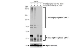
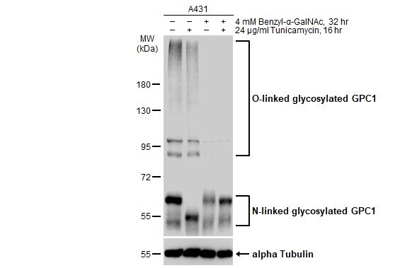
- Western Blot analysis of Glypican 1 was performed by separating 30 µg of untreated (–) and treated (+) A431 whole cell extracts by 7.5% SDS-PAGE. Proteins were transferred to a membrane and probed with a Glypican 1 Polyclonal Antibody (Product # PA5-28055) at a dilution of 1:3000.
- Submitted by
- Invitrogen Antibodies (provider)
- Main image
- Experimental details
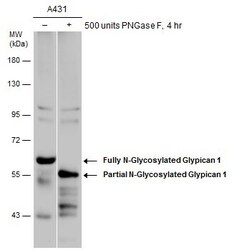
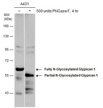
- Western Blot analysis of Glypican 1 was performed by separating 30 µg of untreated (–) and treated (+) A431 whole cell extracts by 7.5% SDS-PAGE. Proteins were transferred to a membrane and probed with a Glypican 1 Polyclonal Antibody (Product # PA5-28055) at a dilution of 1:500.
- Submitted by
- Invitrogen Antibodies (provider)
- Main image
- Experimental details
- Western Blot analysis of Glypican 1 was performed by separating 30 µg of various whole cell extracts by 7.5% SDS-PAGE. Proteins were transferred to a membrane and probed with a Glypican 1 Polyclonal Antibody (Product # PA5-28055) at a dilution of 1:1500.
- Submitted by
- Invitrogen Antibodies (provider)
- Main image
- Experimental details
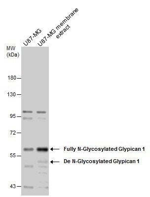
- Western Blot analysis of Glypican 1 was performed by separating 30 µg of U87-MG whole cell and membrane extracts by 7.5% SDS-PAGE. Proteins were transferred to a membrane and probed with a Glypican 1 Polyclonal Antibody (Product # PA5-28055) at a dilution of 1:1500 and a HRP-conjugated anti-rabbit IgG secondary antibody.
Supportive validation
- Submitted by
- Invitrogen Antibodies (provider)
- Main image

- Experimental details
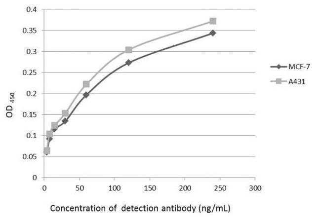
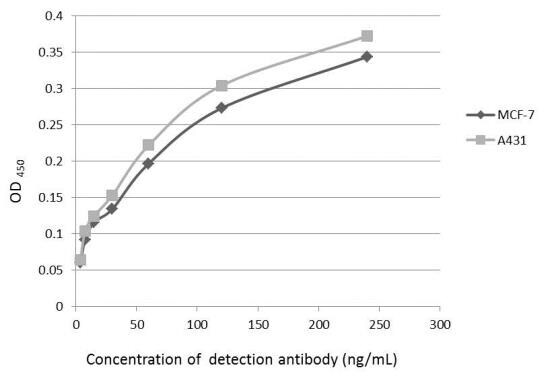
- An ELISA plate is coated with MCF-7 and A431 cells. The coated cells are detected with Glypican 1 antibody (N3C3) (Product # PA5-28055) at concentration ranged from 7.5 to 240 ng/mL.
- Submitted by
- Invitrogen Antibodies (provider)
- Main image

- Experimental details
- An ELISA plate is coated with MCF-7 and A431 cells. The coated cells are detected with Glypican 1 Polyclonal Antibody (Product # PA5-28055) at concentration ranged from 7.5 to 240 ng/mL.
Supportive validation
- Submitted by
- Invitrogen Antibodies (provider)
- Main image
- Experimental details
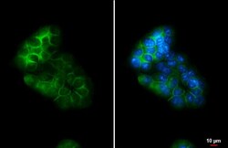
- Immunocytochemistry-Immunofluorescence analysis of Glypican 1 was performed in MCF 7 cells fixed in ice cold MeOH for 5 min. Green: Glypican 1 Polyclonal Antibody (Product # PA5 28055) diluted at 1:500. Blue: Hoechst 33342 staining. Scale bar = 10 µm.

Supportive validation
- Submitted by
- Invitrogen Antibodies (provider)
- Main image
- Experimental details
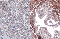
- Immunohistochemistry (Paraffin) analysis of Glypican 1 was performed in paraffin-embedded human normal pancreas (left) and pancreatic cancer (right) tissue using Glypican 1 Polyclonal Antibody (Product # PA5-28055) at a dilution of 1:500. Antigen Retrieval: Citrate buffer, pH 6.0, 15 min.

- Submitted by
- Invitrogen Antibodies (provider)
- Main image

- Experimental details
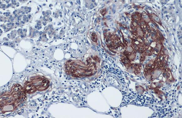
- Glypican 1 Polyclonal Antibody detects Glypican 1 protein at cell membrane and cytoplasm by immunohistochemical analysis. Sample: Paraffin-embedded human pancreatic cancer. Glypican 1 stained by Glypican 1 Polyclonal Antibody (Product # PA5-28055) diluted at 1:1,000. Antigen Retrieval: Citrate buffer, pH 6.0, 15 min.
Supportive validation
- Submitted by
- Invitrogen Antibodies (provider)
- Main image

- Experimental details
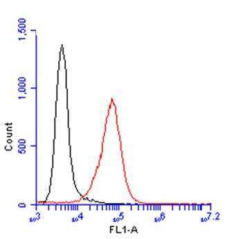
- Flow Cytometry analysis of TLR3 was performed in A431 cells using TLR3 Monoclonal Antibody (40C1285) (Product # MA1-25649) (red) at a dilution of 1:100. Black: Unlabelled sample was used as a control. Acquisition of 20,000 events were collected using a Dylight 488-conjugated secondary antibody for FACS analysis.
Supportive validation
- Submitted by
- Invitrogen Antibodies (provider)
- Main image
- Experimental details
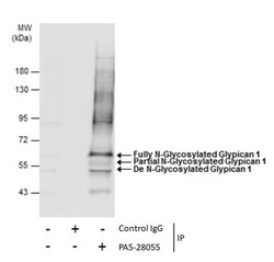
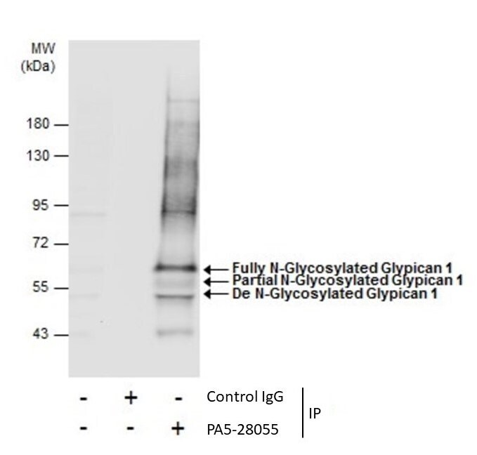
- Immunoprecipitation of Glypican 1 was performed in MCF-7 whole cell extracts using 5 µg of Glypican 1 Polyclonal Antibody (Product # PA5-28055). Samples were transferred to a membrane and probed with Glypican 1 Polyclonal Antibody as a primary antibody and an HRP-conjugated anti-Rabbit IgG was used as a secondary antibody.
- Submitted by
- Invitrogen Antibodies (provider)
- Main image

- Experimental details
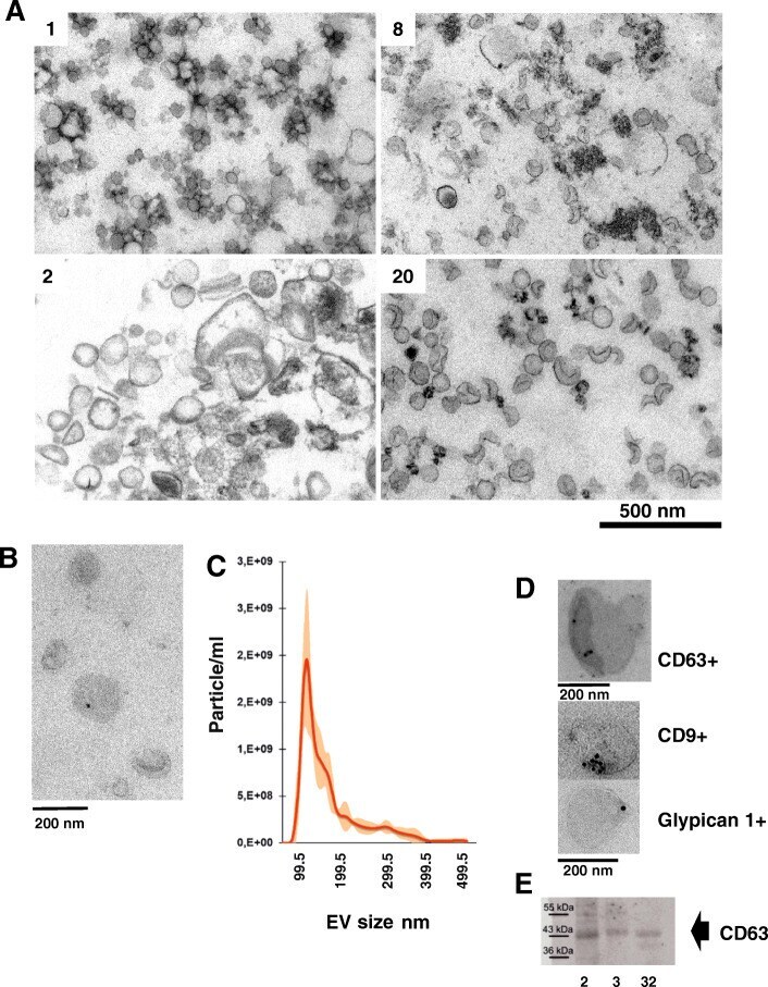
- Fig. 4 Sweat particles. Visual characterization of sweat particles. Panels A : TEM, thin sections of plastic embedded pelleted sweat from 4 donors number corresponding to sequencing library (Table 1 ). B : TEM, negative staining of ExoEasy isolated sweat EVs, C : NTA analysis of Exoeasy EVs, D : TEM, Immunostaining of isolated sweat EVs, E: western blot, protein from Exoeasy sweat preparations were stained with anti-CD63 antibody (ab193349). Full size western blot with region selected marked is shown as supplementary Fig. 6