Explore
 Validate
 Learn
Learn Western blot
Antibody data
- Antibody Data
- Antigen structure
- References [17]
- Comments [0]
- Validations
- Western blot [4]
- Immunocytochemistry [4]
- Immunohistochemistry [3]
- Flow cytometry [3]
- Other assay [1]
Submit
Validation data
Reference
Comment
Report error
- Product number
- MA3-060 - Provider product page

- Provider
- Invitrogen Antibodies
- Product name
- ARF1/ARF3/ARF5/ARF6 Monoclonal Antibody (1D9)
- Antibody type
- Monoclonal
- Antigen
- Recombinant full-length protein
- Description
- MA3-060 detects ADP-ribosylation factor1 (Arf1), Arf3, Arf5 and Arf6 and about ten-fold less well with Arf4 from human, mouse and rat tissues.
- Antibody clone number
- 1D9
- Concentration
- Conc. Not Determined
Submitted references The ARF GAP ELMOD2 acts with different GTPases to regulate centrosomal microtubule nucleation and cytokinesis.
NF-Y inactivation causes atypical neurodegeneration characterized by ubiquitin and p62 accumulation and endoplasmic reticulum disorganization.
Arf GTPase-activating protein ASAP1 interacts with Rab11 effector FIP3 and regulates pericentrosomal localization of transferrin receptor-positive recycling endosome.
ARF1 is directly involved in dynamin-independent endocytosis.
Activation of cellular Arf GTPases by poliovirus protein 3CD correlates with virus replication.
PI4P promotes the recruitment of the GGA adaptor proteins to the trans-Golgi network and regulates their recognition of the ubiquitin sorting signal.
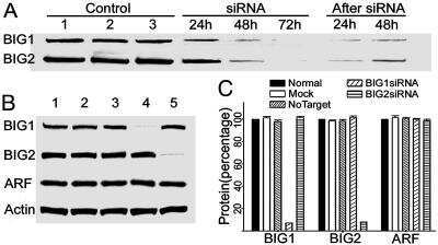
Association of brefeldin A-inhibited guanine nucleotide-exchange protein 2 (BIG2) with recycling endosomes during transferrin uptake.
Arf6 plays an early role in platelet activation by collagen and convulxin.
Poliovirus proteins induce membrane association of GTPase ADP-ribosylation factor.
Inhibition of membrane tubule formation and trafficking by isotetrandrine, an antagonist of G-protein-regulated phospholipase A2 enzymes.
Requirement for Arf6 in breast cancer invasive activities.
Islet cell autoantigen of 69 kDa is an arfaptin-related protein associated with the Golgi complex of insulinoma INS-1 cells.
A role for calcium in stabilizing transport vesicle coats.
An ADP-ribosylation factor GTPase-activating protein Git2-short/KIAA0148 is involved in subcellular localization of paxillin and actin cytoskeletal organization.
Regulation of Golgi structure and function by ARF-like protein 1 (Arl1).
GGAs: a family of ADP ribosylation factor-binding proteins related to adaptors and associated with the Golgi complex.
ADP-Ribosylation factor 1 (ARF1) regulates recruitment of the AP-3 adaptor complex to membranes.
Turn RE, East MP, Prekeris R, Kahn RA
Molecular biology of the cell 2020 Aug 15;31(18):2070-2091
Molecular biology of the cell 2020 Aug 15;31(18):2070-2091
NF-Y inactivation causes atypical neurodegeneration characterized by ubiquitin and p62 accumulation and endoplasmic reticulum disorganization.
Yamanaka T, Tosaki A, Kurosawa M, Matsumoto G, Koike M, Uchiyama Y, Maity SN, Shimogori T, Hattori N, Nukina N
Nature communications 2014 Feb 25;5:3354
Nature communications 2014 Feb 25;5:3354
Arf GTPase-activating protein ASAP1 interacts with Rab11 effector FIP3 and regulates pericentrosomal localization of transferrin receptor-positive recycling endosome.
Inoue H, Ha VL, Prekeris R, Randazzo PA
Molecular biology of the cell 2008 Oct;19(10):4224-37
Molecular biology of the cell 2008 Oct;19(10):4224-37
ARF1 is directly involved in dynamin-independent endocytosis.
Kumari S, Mayor S
Nature cell biology 2008 Jan;10(1):30-41
Nature cell biology 2008 Jan;10(1):30-41
Activation of cellular Arf GTPases by poliovirus protein 3CD correlates with virus replication.
Belov GA, Habbersett C, Franco D, Ehrenfeld E
Journal of virology 2007 Sep;81(17):9259-67
Journal of virology 2007 Sep;81(17):9259-67
PI4P promotes the recruitment of the GGA adaptor proteins to the trans-Golgi network and regulates their recognition of the ubiquitin sorting signal.
Wang J, Sun HQ, Macia E, Kirchhausen T, Watson H, Bonifacino JS, Yin HL
Molecular biology of the cell 2007 Jul;18(7):2646-55
Molecular biology of the cell 2007 Jul;18(7):2646-55
Association of brefeldin A-inhibited guanine nucleotide-exchange protein 2 (BIG2) with recycling endosomes during transferrin uptake.
Shen X, Xu KF, Fan Q, Pacheco-Rodriguez G, Moss J, Vaughan M
Proceedings of the National Academy of Sciences of the United States of America 2006 Feb 21;103(8):2635-40
Proceedings of the National Academy of Sciences of the United States of America 2006 Feb 21;103(8):2635-40
Arf6 plays an early role in platelet activation by collagen and convulxin.
Choi W, Karim ZA, Whiteheart SW
Blood 2006 Apr 15;107(8):3145-52
Blood 2006 Apr 15;107(8):3145-52
Poliovirus proteins induce membrane association of GTPase ADP-ribosylation factor.
Belov GA, Fogg MH, Ehrenfeld E
Journal of virology 2005 Jun;79(11):7207-16
Journal of virology 2005 Jun;79(11):7207-16
Inhibition of membrane tubule formation and trafficking by isotetrandrine, an antagonist of G-protein-regulated phospholipase A2 enzymes.
Chan D, Strang M, Judson B, Brown WJ
Molecular biology of the cell 2004 Apr;15(4):1871-80
Molecular biology of the cell 2004 Apr;15(4):1871-80
Requirement for Arf6 in breast cancer invasive activities.
Hashimoto S, Onodera Y, Hashimoto A, Tanaka M, Hamaguchi M, Yamada A, Sabe H
Proceedings of the National Academy of Sciences of the United States of America 2004 Apr 27;101(17):6647-52
Proceedings of the National Academy of Sciences of the United States of America 2004 Apr 27;101(17):6647-52
Islet cell autoantigen of 69 kDa is an arfaptin-related protein associated with the Golgi complex of insulinoma INS-1 cells.
Spitzenberger F, Pietropaolo S, Verkade P, Habermann B, Lacas-Gervais S, Mziaut H, Pietropaolo M, Solimena M
The Journal of biological chemistry 2003 Jul 11;278(28):26166-73
The Journal of biological chemistry 2003 Jul 11;278(28):26166-73
A role for calcium in stabilizing transport vesicle coats.
Ahluwalia JP, Topp JD, Weirather K, Zimmerman M, Stamnes M
The Journal of biological chemistry 2001 Sep 7;276(36):34148-55
The Journal of biological chemistry 2001 Sep 7;276(36):34148-55
An ADP-ribosylation factor GTPase-activating protein Git2-short/KIAA0148 is involved in subcellular localization of paxillin and actin cytoskeletal organization.
Mazaki Y, Hashimoto S, Okawa K, Tsubouchi A, Nakamura K, Yagi R, Yano H, Kondo A, Iwamatsu A, Mizoguchi A, Sabe H
Molecular biology of the cell 2001 Mar;12(3):645-62
Molecular biology of the cell 2001 Mar;12(3):645-62
Regulation of Golgi structure and function by ARF-like protein 1 (Arl1).
Lu L, Horstmann H, Ng C, Hong W
Journal of cell science 2001 Dec;114(Pt 24):4543-55
Journal of cell science 2001 Dec;114(Pt 24):4543-55
GGAs: a family of ADP ribosylation factor-binding proteins related to adaptors and associated with the Golgi complex.
Dell'Angelica EC, Puertollano R, Mullins C, Aguilar RC, Vargas JD, Hartnell LM, Bonifacino JS
The Journal of cell biology 2000 Apr 3;149(1):81-94
The Journal of cell biology 2000 Apr 3;149(1):81-94
ADP-Ribosylation factor 1 (ARF1) regulates recruitment of the AP-3 adaptor complex to membranes.
Ooi CE, Dell'Angelica EC, Bonifacino JS
The Journal of cell biology 1998 Jul 27;142(2):391-402
The Journal of cell biology 1998 Jul 27;142(2):391-402
No comments: Submit comment
Supportive validation
- Submitted by
- Invitrogen Antibodies (provider)
- Main image

- Experimental details
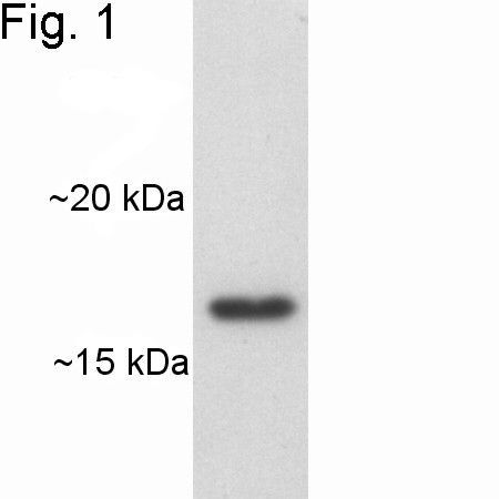
- Western blot of Arf on canine heart extract using Product # MA3-060.
- Submitted by
- Invitrogen Antibodies (provider)
- Main image

- Experimental details
- Western blot of Arf on canine heart extract using Product # MA3-060.

- Submitted by
- Invitrogen Antibodies (provider)
- Main image
- Experimental details
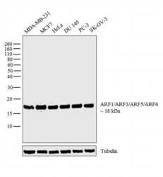
- Western blot analysis was performed on whole cell extracts (30 µg lysate) of MDA-MB-231 (Lane 1), MCF7 (Lane 2), HeLa (Lane 3), DU 145 (Lane 4), PC-3 (Lane 5) and SK-OV-3 (Lane 6). The blot was probed with Anti-ARF1/ARF3/ARF5/ARF6 Monoclonal Antibody (Product # MA3-060, 1:3000 dilution) and detected by chemiluminescence using Goat anti-Mouse IgG (H+L) Superclonal™ Secondary Antibody, HRP conjugate (Product # A28177, 0.25 µg/ml, 1:4000 dilution). A 18 kDa band corresponding to ARF1/ARF3/ARF5/ARF6 was observed across the cell lines tested.

- Submitted by
- Invitrogen Antibodies (provider)
- Main image

- Experimental details
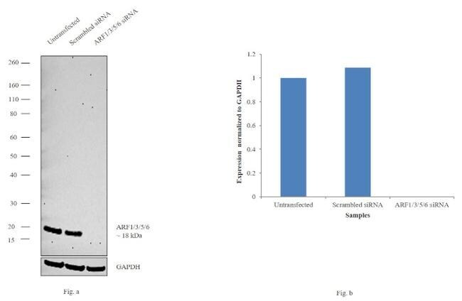
- Knockdown of ARF1/ARF3/ARF5/ARF6 was achieved by transfecting HeLa with ARF1/ARF3/ARF5/ARF6 specific siRNAs (Silencer® select Product # s1555, s1552 ). Western blot analysis (Fig. a) was performed using whole cell extracts from the ARF1/ARF3/ARF5/ARF6 knockdown cells (lane 3), non-specific scrambled siRNA transfected cells (lane 2) and untransfected cells (lane 1). The blot was probed with ARF1/ARF3/ARF5/ARF6 Monoclonal Antibody (Product # MA3-060, 1:500 dilution) and Goat anti-Mouse IgG (H+L) Superclonal™ Secondary Antibody, HRP conjugate (Product # A28177, 0.25µg/ml, 1:4000 dilution). Densitometric analysis of this western blot is shown in histogram (Fig. b). Decrease in signal upon siRNA mediated knock down confirms that antibody is specific to ARF1/ARF3/ARF5/ARF6.
Supportive validation
- Submitted by
- Invitrogen Antibodies (provider)
- Main image
- Experimental details
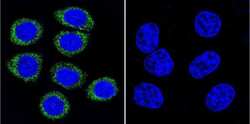
- Immunofluorescent analysis of ADP-Ribosylation Factor using ADP-Ribosylation Factor Monoclonal Antibody (1D9) (Product # MA3-060) shows staining in Hela Cells. ADP-Ribosylation Factor (green), F-Actin staining with Phalloidin (red) and nuclei with DAPI (blue) is shown. Cells were grown on chamber slides and fixed with formaldehyde prior to staining. Cells were probed without (control) or with an antibody recognizing ADP-Ribosylation Factor (Product # MA3-060) at a dilution of 1:100 over night at 4 °C, washed with PBS and incubated with a DyLight-488 conjugated secondary antibody (Product # 35552 for GAR, Product # 35503 for GAM). Images were taken at 60X magnification.

- Submitted by
- Invitrogen Antibodies (provider)
- Main image
- Experimental details
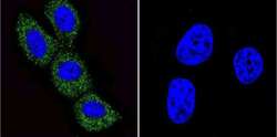
- Immunofluorescent analysis of ADP-Ribosylation Factor using ADP-Ribosylation Factor Monoclonal Antibody (1D9) (Product # MA3-060) shows staining in MCF-7 Cells. ADP-Ribosylation Factor (green), F-Actin staining with Phalloidin (red) and nuclei with DAPI (blue) is shown. Cells were grown on chamber slides and fixed with formaldehyde prior to staining. Cells were probed without (control) or with an antibody recognizing ADP-Ribosylation Factor (Product # MA3-060) at a dilution of 1:100 over night at 4 °C, washed with PBS and incubated with a DyLight-488 conjugated secondary antibody (Product # 35552 for GAR, Product # 35503 for GAM). Images were taken at 60X magnification.
- Submitted by
- Invitrogen Antibodies (provider)
- Main image
- Experimental details
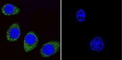
- Immunofluorescent analysis of ADP-Ribosylation Factor using ADP-Ribosylation Factor Monoclonal Antibody (1D9) (Product # MA3-060) shows staining in U251 Cells. ADP-Ribosylation Factor (green), F-Actin staining with Phalloidin (red) and nuclei with DAPI (blue) is shown. Cells were grown on chamber slides and fixed with formaldehyde prior to staining. Cells were probed without (control) or with an antibody recognizing ADP-Ribosylation Factor (Product # MA3-060) at a dilution of 1:100 over night at 4 °C, washed with PBS and incubated with a DyLight-488 conjugated secondary antibody (Product # 35552 for GAR, Product # 35503 for GAM). Images were taken at 60X magnification.

- Submitted by
- Invitrogen Antibodies (provider)
- Main image
- Experimental details
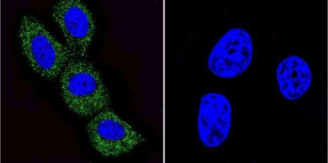
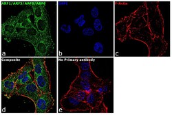
- Immunofluorescence analysis of ARF1/ARF3/ARF5/ARF6 was performed using 70% confluent log phase MDA-MB-231 cells. The cells were fixed with 4% paraformaldehyde for 10 minutes, permeabilized with 0.1% Triton™ X-100 for 15 minutes, and blocked with 1% BSA for 1 hour at room temperature. The cells were labeled with ARF1/ARF3/ARF5/ARF6 Mouse Monoclonal Antibody (Product # MA3-060) at 1:100 dilution in 0.1% BSA, incubated at 4 degree Celsius overnight and then labeled with Goat anti-Mouse IgG (H+L) Superclonal™ Secondary Antibody, Alexa Fluor® 488 conjugate (Product # A28175) at a dilution of 1:2000 for 45 minutes at room temperature (Panel a: green). Nuclei (Panel b: blue) were stained with ProLong™ Diamond Antifade Mountant with DAPI (Product # P36962). F-actin (Panel c: red) was stained with Rhodamine Phalloidin (Product # R415, 1:300). Panel d represents the merged image showing cytoplasmic localization. Panel e represents control cells with no primary antibody to assess background. The images were captured at 60X magnification.

Supportive validation
- Submitted by
- Invitrogen Antibodies (provider)
- Main image

- Experimental details
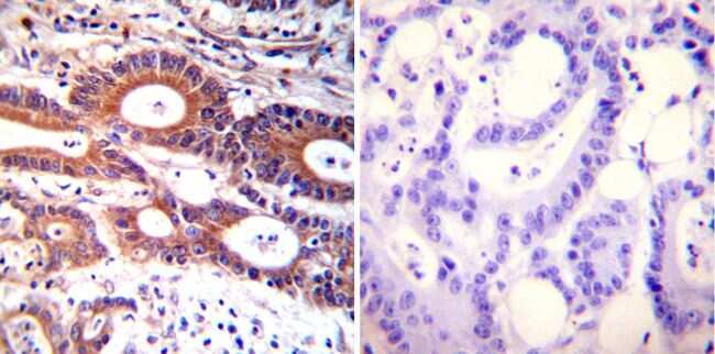
- Immunohistochemistry was performed on cancer biopsies of deparaffinized Human colon carcinoma tissues. To expose target proteins, heat induced antigen retrieval was performed using 10mM sodium citrate (pH6.0) buffer, microwaved for 8-15 minutes. Following antigen retrieval tissues were blocked in 3% BSA-PBS for 30 minutes at room temperature. Tissues were then probed at a dilution of 1:100 with a mouse monoclonal antibody recognizing ADP-Ribosylation Factor (Product # MA3-060) or without primary antibody (negative control) overnight at 4°C in a humidified chamber. Tissues were washed extensively with PBST and endogenous peroxidase activity was quenched with a peroxidase suppressor. Detection was performed using a biotin-conjugated secondary antibody and SA-HRP, followed by colorimetric detection using DAB. Tissues were counterstained with hematoxylin and prepped for mounting.
- Submitted by
- Invitrogen Antibodies (provider)
- Main image

- Experimental details
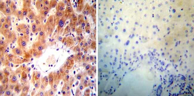
- Immunohistochemistry was performed on normal deparaffinized Human liver tissue tissues. To expose target proteins, heat induced antigen retrieval was performed using 10mM sodium citrate (pH6.0) buffer, microwaved for 8-15 minutes. Following antigen retrieval tissues were blocked in 3% BSA-PBS for 30 minutes at room temperature. Tissues were then probed at a dilution of 1:50 with a mouse monoclonal antibody recognizing ADP-Ribosylation Factor (Product # MA3-060) or without primary antibody (negative control) overnight at 4°C in a humidified chamber. Tissues were washed extensively with PBST and endogenous peroxidase activity was quenched with a peroxidase suppressor. Detection was performed using a biotin-conjugated secondary antibody and SA-HRP, followed by colorimetric detection using DAB. Tissues were counterstained with hematoxylin and prepped for mounting.
- Submitted by
- Invitrogen Antibodies (provider)
- Main image

- Experimental details
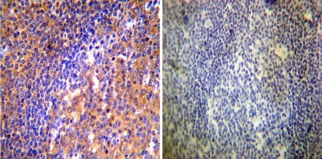
- Immunohistochemistry was performed on normal deparaffinized Human tonsil tissue tissues. To expose target proteins, heat induced antigen retrieval was performed using 10mM sodium citrate (pH6.0) buffer, microwaved for 8-15 minutes. Following antigen retrieval tissues were blocked in 3% BSA-PBS for 30 minutes at room temperature. Tissues were then probed at a dilution of 1:50 with a mouse monoclonal antibody recognizing ADP-Ribosylation Factor (Product # MA3-060) or without primary antibody (negative control) overnight at 4°C in a humidified chamber. Tissues were washed extensively with PBST and endogenous peroxidase activity was quenched with a peroxidase suppressor. Detection was performed using a biotin-conjugated secondary antibody and SA-HRP, followed by colorimetric detection using DAB. Tissues were counterstained with hematoxylin and prepped for mounting.
Supportive validation
- Submitted by
- Invitrogen Antibodies (provider)
- Main image
- Experimental details
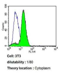
- Flow cytometry analysis of ADP-Ribosylation Factor in 3T3 cells compared to an isotype control (blue). Cells were harvested, adjusted to a concentration of 1-5x10^6 cells/mL, fixed with 2% paraformaldehyde, washed with PBS, and incubated with ADP-Ribosylation Factor monoclonal antibody (Product # MA3-060) at a dilution of 1:80 for 60 min at room temperature. Cells were then blocked in a solution of 2% BSA-PBS for 30 min at room temperature, incubated for 40 min at room temperature in the dark using a Dylight 488-conjugated goat anti-mouse IgG (H+L) secondary antibody, and re-suspended in PBS for FACS analysis.

- Submitted by
- Invitrogen Antibodies (provider)
- Main image

- Experimental details
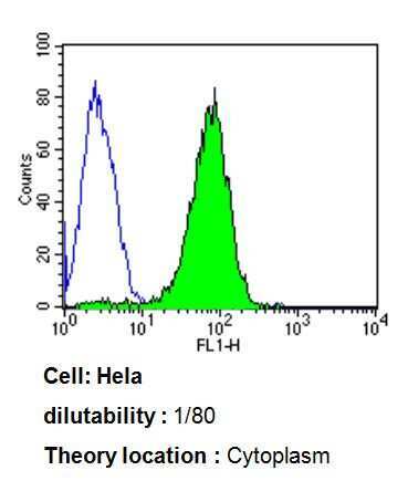
- Flow cytometry analysis of ADP-Ribosylation Factor in Hela cells compared to an isotype control (blue). Cells were harvested, adjusted to a concentration of 1-5x10^6 cells/mL, fixed with 2% paraformaldehyde, washed with PBS, and incubated with ADP-Ribosylation Factor monoclonal antibody (Product # MA3-060) at a dilution of 1:80 for 60 min at room temperature. Cells were then blocked in a solution of 2% BSA-PBS for 30 min at room temperature, incubated for 40 min at room temperature in the dark using a Dylight 488-conjugated goat anti-mouse IgG (H+L) secondary antibody, and re-suspended in PBS for FACS analysis.
- Submitted by
- Invitrogen Antibodies (provider)
- Main image
- Experimental details
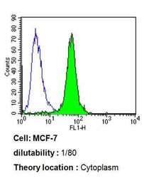
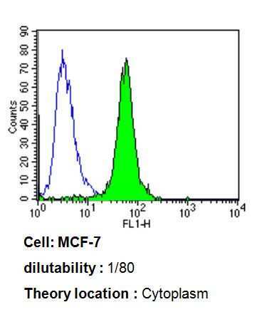
- Flow cytometry analysis of ADP-Ribosylation Factor in MCF-7 cells compared to an isotype control (blue). Cells were harvested, adjusted to a concentration of 1-5x10^6 cells/mL, fixed with 2% paraformaldehyde, washed with PBS, and incubated with ADP-Ribosylation Factor monoclonal antibody (Product # MA3-060) at a dilution of 1:80 for 60 min at room temperature. Cells were then blocked in a solution of 2% BSA-PBS for 30 min at room temperature, incubated for 40 min at room temperature in the dark using a Dylight 488-conjugated goat anti-mouse IgG (H+L) secondary antibody, and re-suspended in PBS for FACS analysis.
Supportive validation
- Submitted by
- Invitrogen Antibodies (provider)
- Main image

- Experimental details
- NULL