 Explore
 Validate
 Learn
LearnA303-504A
antibody from Invitrogen Antibodies
Targeting: TAF1
BA2R, CCG1, CCGS, DYT3, DYT3/TAF1, KAT4, NSCL2, TAF2A, TAFII250
 Western blot
 Immunoprecipitation
Antibody data
- Antibody Data
- Antigen structure
- References [0]
- Comments [0]
- Validations
- Western blot [2]
- Immunohistochemistry [3]
- Other assay [1]
Submit
Validation data
Reference
Comment
Report error
- Product number
- A303-504A - Provider product page

- Provider
- Invitrogen Antibodies
- Product name
- TAF1 Polyclonal Antibody
- Antibody type
- Polyclonal
- Antigen
- Other
- Reactivity
- Human
- Host
- Rabbit
- Isotype
- IgG
- Vial size
- 100 µL
- Concentration
- 1 mg/mL
- Storage
- 4° C
No comments: Submit comment
Supportive validation
- Submitted by
- Invitrogen Antibodies (provider)
- Main image
- Experimental details
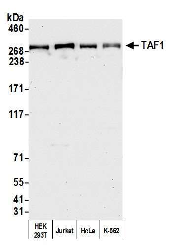
- Detection of human TAF1 by western blot. Samples: Whole cell lysate (50 µg) from HEK293T, Jurkat, HeLa, and K-562 cells prepared using NETN lysis buffer. Antibody: Affinity purified rabbit anti-TAF1 antibody (Product # A303-504A lot 3) used for WB at 0.04 µg/mL. Detection: Chemiluminescence with an exposure time of 30 seconds.
- Submitted by
- Invitrogen Antibodies (provider)
- Main image

- Experimental details
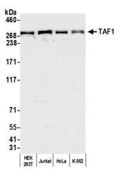
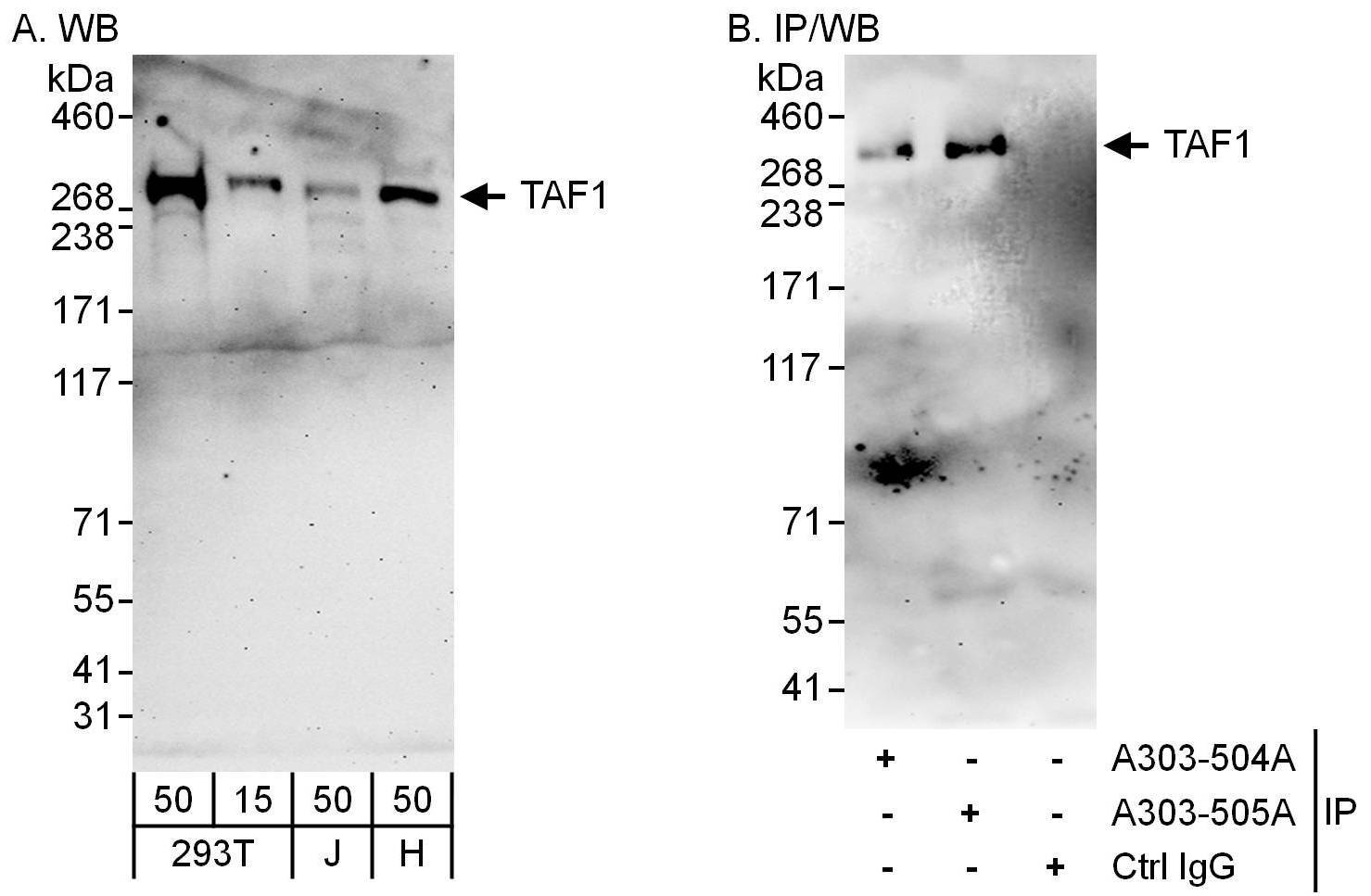
- Detection of human TAF1 by western blot and immunoprecipitation. Samples: Whole cell lysate from 293T (15 and 50 µg for WB; 1 mg for IP, 20% of IP loaded), Jurkat (J; 50 µg) and HeLa (H; 50 µg) cells. Antibodies: Affinity purified rabbit anti-TAF1 antibody A303-504A used for WB at 0.4 µg/ml (A) and 1 µg/ml (B) and used for IP at 6 µg/mg lysate. TAF1 was also immunoprecipitated by rabbit anti-TAF1 antibody A303-505A, which recognizes a downstream epitope. Detection: Chemiluminescence with exposure times of 3 minutes (A and B).
Supportive validation
- Submitted by
- Invitrogen Antibodies (provider)
- Main image
- Experimental details
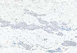
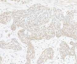
- Detection of human TAF1 by immunohistochemistry. Sample: FFPE section of bladder carcinoma. Antibody: Rabbit anti-TAF1 antibody (Product # A303-504A; lot 3) used at 1:1000 (1 µg/mL). Secondary: HRP-conjugated goat anti-rabbit IgG (Product # A120-501P). Substrate: DAB.

- Submitted by
- Invitrogen Antibodies (provider)
- Main image
- Experimental details
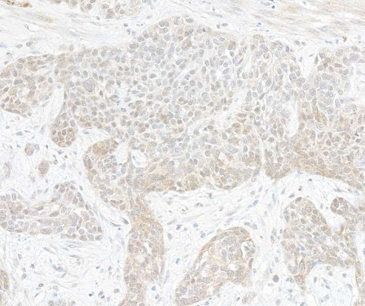
- Detection of human TAF1 by immunohistochemistry. Sample: FFPE section of lung carcinoma. Antibody: Rabbit anti-TAF1 antibody (Product # A303-504A lot 3) used at 1:5000 (0.2 µg/mL). Secondary: HRP-conjugated goat anti-rabbit IgG (A120-501P). Substrate: DAB.
- Submitted by
- Invitrogen Antibodies (provider)
- Main image
- Experimental details
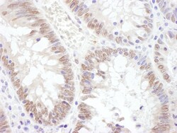
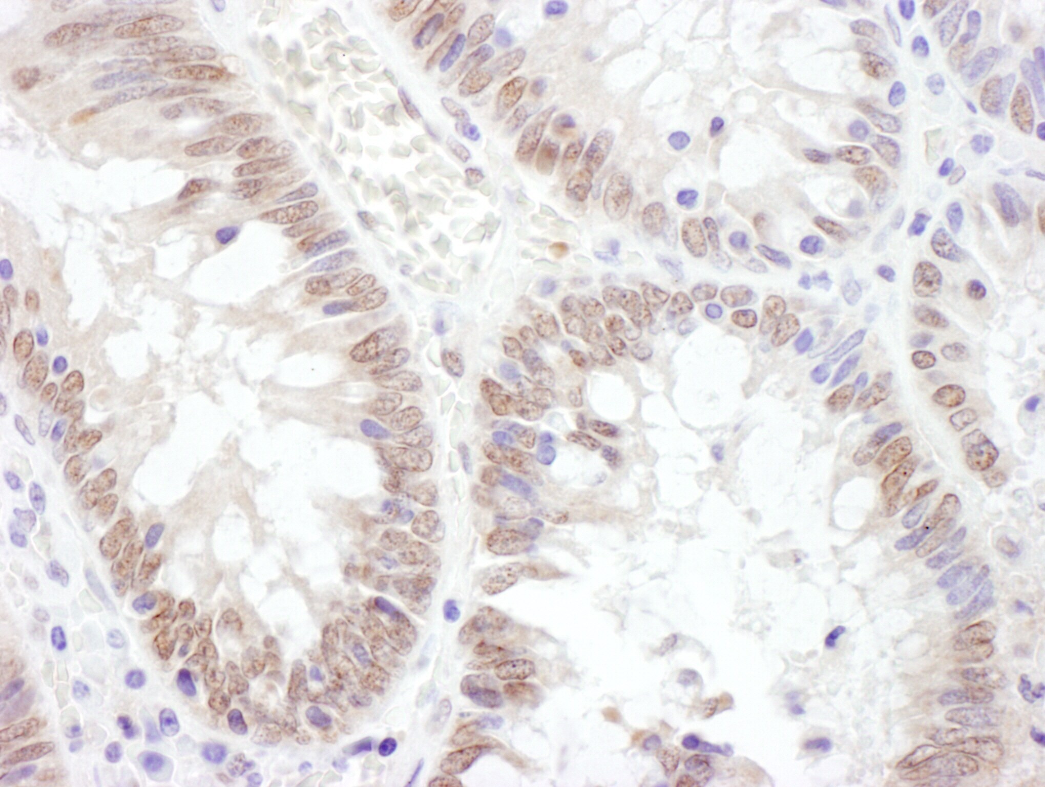
- Detection of human TAF1 by immunohistochemistry. Sample: FFPE section of human colon carcinoma. Antibody: Affinity purified rabbit anti- TAF1 (Cat. No. A303-504A Lot1) used at a dilution of 1:1,000 (1µg/ml). Detection: DAB.
Supportive validation
- Submitted by
- Invitrogen Antibodies (provider)
- Main image
- Experimental details
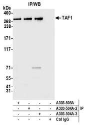
- Detection of human TAF1 by western blot of immunoprecipitates. Samples: Whole cell lysate (1.0 mg per IP reaction; 20% of IP loaded) from HEK293T cells prepared using NETN lysis buffer. Antibodies: Affinity purified rabbit anti-TAF1 antibody (Product # A303-504A lot 3) used for IP at 6 µg per reaction. TAF1 was also immunoprecipitated by a previous lot of this antibody (A303-504A lot 2) and rabbit anti-TAF1 antibody (Product # A303-505A). For blotting immunoprecipitated TAF1 (Product # A303-504A) was used at 0.04 µg/mL. Detection: Chemiluminescence with an exposure time of 3 seconds.
